
Presentation
History of longstanding slow-growing, painless, vulvar mass.
Patient Data
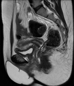

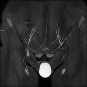
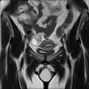
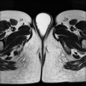
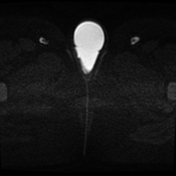
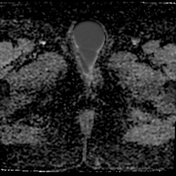
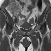
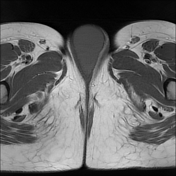

Giant vulvar cystic lesion which elicits low signal on T1 WI, high signal on T2 WI with diffusion restriction and low ADC values. There is no suspicious solid component and no deep extension.
Case Discussion
This is an uncommon case of a giant vulvar epidermoid cyst. The longstanding history, external location and diffusion restriction were the main clues for the diagnosis. The aetiology is unknown, but might be attributed to previous genital mutilation which is common in this culture.
Vulvar epidermoid cysts are uncommon and usually occur secondary to vulvar trauma or surgical interventions including female genital mutilation in some cultures and episiotomy.
MRI is helpful for characterisation, localisation of the lesion and detection of deep perineal extension.


 Unable to process the form. Check for errors and try again.