
Presentation
Dysarthria and left sided weakness
Patient Data
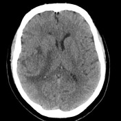

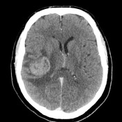

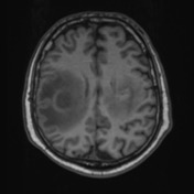
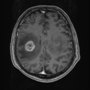
3.4 cm right frontoparietal enhancing lesion with marked vasogenic oedema and regional positive mass effect. 3.2 cm left frontoparietal partially calcified lesion with minor adjacent vasogenic oedema and regional sulcal effacement. An additional smaller enhancing focus at the left superior frontal gyrus which measures 1.6 cm. 4 mm of midline shift to the left.
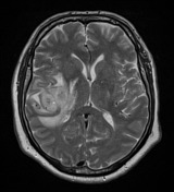

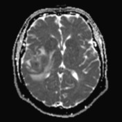
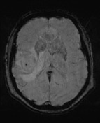
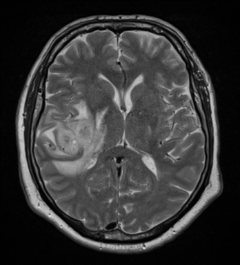
Mass in the right inferior frontal gyrus extending into the white matter of the right corona radiata. Thick peripheral nodular enhancement, and central non enhancement. Central reduction of ADC value. Extensive surrounding oedema. Ill-defined infiltrating mass in the left centrum semiovale. Intrinsic right T1 signal. No convincing enhancement. Surrounding T2 and FLAIR hyperintense white matter abnormality extends into the left pre and post central gyri, both of which are expanded. Intralesional calcification. Multiple additional small foci of ring enhancement. 7 mm of midline shift to the left. No hydrocephalus.
CLINICAL NOTES: (R) frontal lesion
MACROSCOPIC DESCRIPTION: 1. "Brain tissue": A piece of firm tan tissue
FROZEN SECTION DIAGNOSIS: High grade glioma. A1.
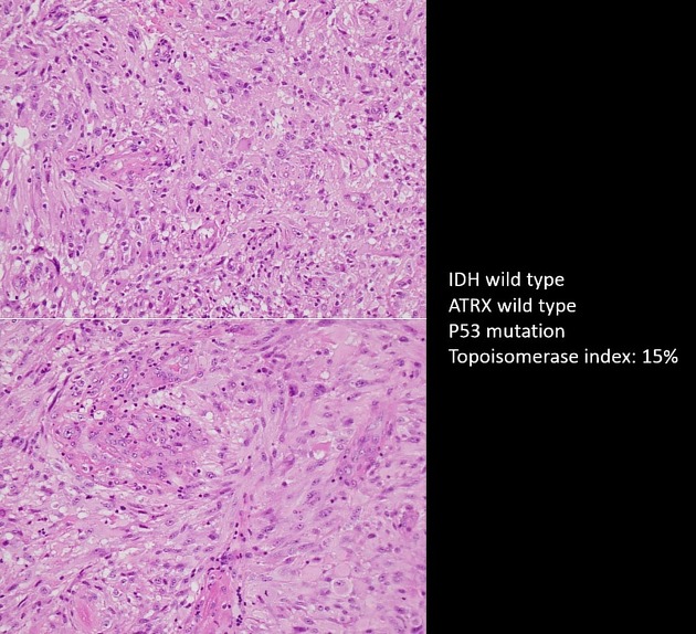
MICROSCOPIC DESCRIPTION: 1-3. Sections show a moderately cellular astrocytic glioma with extensive areas of tumour necrosis. There are multiple foci of microvascular proliferation. Tumour cells demonstrate marked nuclear pleomorphism with hyperchromasia and prominent nucleoli. Frequent mitoses are observed.
IMMUNOHISTOCHEMISTRY: GFAP positive Nestin positive (high) Nogo A negative IDH-1 R132H negative (not mutated) ATRX positive (not mutated) P53 positive P16 negative Topoisomerase labelling index 15%.
DIAGNOSIS: The features are of glioblastoma IDH wild type (WHO Grade IV)


 Unable to process the form. Check for errors and try again.