Glioblastoma NOS
Updates to Case Attributes
Final Diagnosis:
Brain, left frontal: Astrocytoma grade 4/4 (glioblastoma) using the Daumas-Duport grading system
Histology
Microscopic Description:
Sections show the specimen to consist predominantly of necrotic tissue, in areas containing viable glioma that is high grade. The tumortumour cells display small cell morphologic characteristics, possible oligodendroglial features, and astrocytic elements with a fibrillar appearance. Mitotic figures are identified. Vascular proliferation is observed, including glomeruloid aggregates of proliferating capillaries. As previously mentioned, tumortumour necrosis is extensive. Abnormally large ectatic vascular structures are also seen, and are consistent with a vascular malformation. Several vessels are thrombosed.
Final diagnosis: glioblastoma
Note: This case predates the recent (2016) revision WHO classification of CNS tumours and IDH status is not available. As such, this tumour would now be classified as a glioblastoma NOS.
-<p>Final Diagnosis:</p><p>Brain, left frontal: Astrocytoma grade 4/4 (<a href="/articles/glioblastoma">glioblastoma</a>) using the Daumas-Duport grading system.</p><p>Microscopic Description:</p><p>Sections show the specimen to consist predominantly of necrotic tissue, in areas containing viable glioma that is high grade. The tumor cells display small cell morphologic characteristics, possible oligodendroglial features, and astrocytic elements with a fibrillar appearance. Mitotic figures are identified. Vascular proliferation is observed, including glomeruloid aggregates proliferating capillaries. As previously mentioned, tumor necrosis is extensive. Abnormally large ectatic vascular structures are also seen, and are consistent with a vascular malformation. Several vessels are thrombosed.</p>- +<p> </p><p>The patient went on to have surgery. </p><p><strong>Histology</strong></p><p>Microscopic Description:</p><p>Sections show the specimen to consist predominantly of necrotic tissue, in areas containing viable glioma that is high grade. The tumour cells display small cell morphologic characteristics, possible oligodendroglial features, and astrocytic elements with a fibrillar appearance. Mitotic figures are identified. Vascular proliferation is observed, including glomeruloid aggregates of proliferating capillaries. As previously mentioned, tumour necrosis is extensive. Abnormally large ectatic vascular structures are also seen and are consistent with a vascular malformation. Several vessels are thrombosed.</p><p>Final diagnosis: <a title="Glioblastoma" href="/articles/glioblastoma">glioblastoma</a></p><p><strong>Note:</strong> This case predates the recent (2016) revision WHO classification of CNS tumours and IDH status is not available. As such, this tumour would now be classified as a <a title="Glioblastoma NOS" href="/articles/glioblastoma-nos">glioblastoma NOS</a>.</p>
Updates to Study Attributes
PendingA larger left frontal lobe peripherally enhancing mass with central non-enhancement (suggesting necrosis) is present. It is surrounded by oedema and exerts positive mass effect.
Image MRI (T1 C+) ( update )
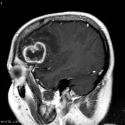
Image MRI (T1 C+) ( update )
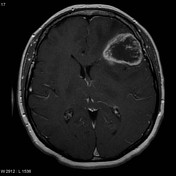
Image MRI (T1) ( update )

Image MRI (T1 C+) ( update )
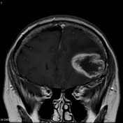
Image MRI (FLAIR) ( update )

Image MRI (DWI) ( update )
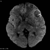
Image MRI (T2) ( update )
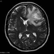
Image 2 MRI (T1 C+) ( update )
Image 3 MRI (FLAIR) ( update )
Image 4 MRI (T2) ( update )
Image 5 MRI (DWI) ( update )
Image 6 MRI (T1 C+) ( update )
Image 7 MRI (T1 C+) ( update )