Glioblastoma NOS
Updates to Study Attributes
Updates to Study Attributes
Pending.
Image 2 MRI (T1 C+) ( update )
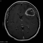
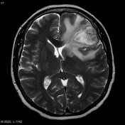
Image 4 MRI (T2) ( update )
Updates to Case Attributes
Final Diagnosis:
Brain, left frontal: Astrocytoma grade 4/4 (glioblastoma multiforme) using the Daumas-Duport grading system.
Microscopic Description:
Sectionsshow the specimen to consist predominantly of necrotic tissue, in areascontaining viable glioma that is high grade. The tumor cells displaysmall cell morphologic characteristics, possible oligodendroglialfeatures, and astrocytic elements with a fibrillar appearance. Mitoticfigures are identified. Vascular proliferation is observed, includingglomeruloid aggregates proliferating capillaries. As previouslymentioned, tumor necrosis is extensive. Abnormally large ectaticvascular structures are also seen, and are consistent with a vascularmalformation. Several vessels are thrombosed.
-<p>Final Diagnosis:</p><p>Brain, left frontal: Astrocytoma grade 4/4 (<a href="/articles/glioblastoma-multiforme">glioblastoma multiforme</a>) using the Daumas-Duport grading system.</p><p>Microscopic Description:</p><p>Sections-show the specimen to consist predominantly of necrotic tissue, in areas-containing viable glioma that is high grade. The tumor cells display-small cell morphologic characteristics, possible oligodendroglial-features, and astrocytic elements with a fibrillar appearance. Mitotic-figures are identified. Vascular proliferation is observed, including-glomeruloid aggregates proliferating capillaries. As previously-mentioned, tumor necrosis is extensive. Abnormally large ectatic-vascular structures are also seen, and are consistent with a vascular-malformation. Several vessels are thrombosed.</p>- +<p>Final Diagnosis:</p><p>Brain, left frontal: Astrocytoma grade 4/4 (<a href="/articles/glioblastoma">glioblastoma</a>) using the Daumas-Duport grading system.</p><p>Microscopic Description:</p><p>Sections show the specimen to consist predominantly of necrotic tissue, in areas containing viable glioma that is high grade. The tumor cells display small cell morphologic characteristics, possible oligodendroglial features, and astrocytic elements with a fibrillar appearance. Mitotic figures are identified. Vascular proliferation is observed, including glomeruloid aggregates proliferating capillaries. As previously mentioned, tumor necrosis is extensive. Abnormally large ectatic vascular structures are also seen, and are consistent with a vascular malformation. Several vessels are thrombosed.</p>
Tags changed:
- findings