
Presentation
History of a progressive headache. Diagnosis confirmed by biopsy. Patient in follow-up, with previous radiotherapy.
Patient Data
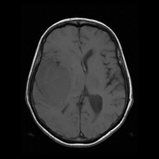

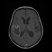
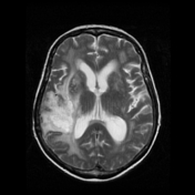
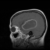

MRI demonstrates an intra-axial mass involving the right temporal lobe, predominantly hypointense on T1 and hyperintense on T2, exhibiting a heterogeneous and peripheral enhancement in the post-contrast study.
Midline shift, cerebral oedema and marked mass effect, especially upon the atrium, are noted.
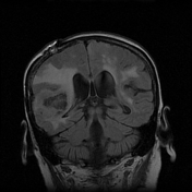
Also, hyperintense areas involving supratentorial white matter on T2 and FLAIR weighted imaging, probably secondary to microangiopathy and/or radiotherapy.
Case Discussion
The patient has had the diagnosis confirmed by biopsy and histology demonstrated a glioblastoma but with unavailable IDH mutation status, then this tumour has been classified as a glioblastoma NOS.


 Unable to process the form. Check for errors and try again.