
Presentation
Right upper abdominal pain.
Patient Data


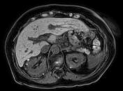
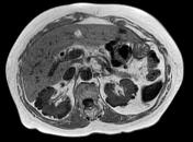
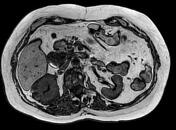
CT abdomen after intake of oral contrast, without injected contrast due to renal failure.
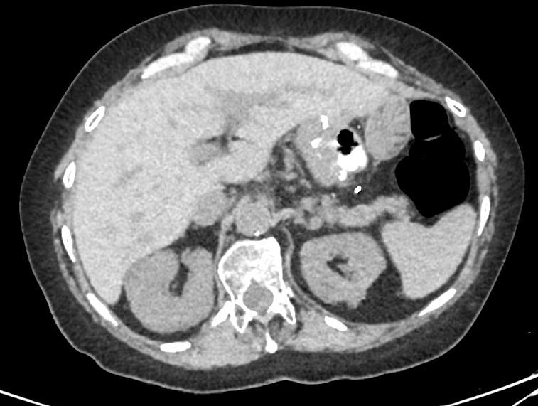
Retained contrast material in the oesophagus. Substantial focal wall thickening in the distal oesophagus.
Status post Roux-en-Y gastric bypass.
Status post cholecystectomy.
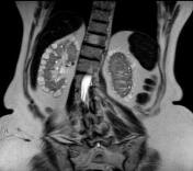
Large diverticulum in 2nd duodenal segment.
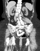
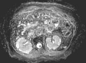
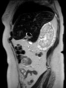
Innumerable small cortical renal cysts envelop the kidneys, seen more clearly around the right kidney. Most cysts are subcentimetre in diameter. Several high-density cysts in the right kidney.
Lumbar scoliosis convex to the right.
Non-contrast MRI done 3.5 years later - suspected "complex renal cyst".
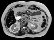
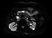
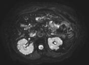
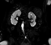

Only non-contrast scans done due to renal failure.
Normal-sized kidneys without hydronephrosis. Each kidney contains innumerable small subcapsular cortical cysts surrounding the periphery, some filled with or containing a sediment of proteinaceous material.
Haemangiomas in several vertebrae.
Case Discussion
Features pathognomonic of glomerulocystic kidney disease.


 Unable to process the form. Check for errors and try again.