
Presentation
Pronounced bilateral proptosis and preseptal soft tissue swelling and orbital pain with a history of long-standing graves disease.
Patient Data


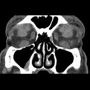
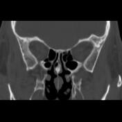
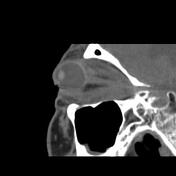
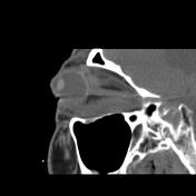
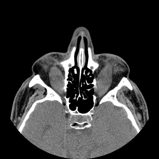
Bilateral pronounced proptosis, enlargement of all extraocular muscles containing hypodense foci, enlarged and prolapsed lacrimal glands, and remodelling of orbital medial walls due to enlarged medial rectus muscles on both sides are seen. Crowding of the apex of orbits is noted. Enlarged orbital fat with bulging within eyelids is seen. Preseptal soft tissue swelling more in lower eyelid is also noted.
Case Discussion
The case illustrates a relatively rare enlargement of all extraocular muscles containing hypodense foci on both sides of orbital cavities in Graves disease. The hypodense foci in extraocular muscles are usually due to mucopolysaccharides accumulation.


 Unable to process the form. Check for errors and try again.