
Presentation
Progressive painless swelling of the right buccal cheek of 8 years duration.
Patient Data
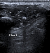
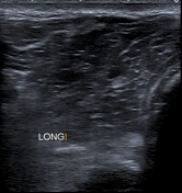
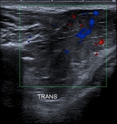

The ultrasound images show an ill-defined hypoechoic mass within the right buccal space of heterogeneous echotexture with cystic spaces within as well as foci of calcification (phleboliths). The color Doppler shows an arterial and venous flow within the mass.
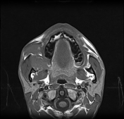


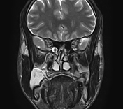
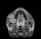
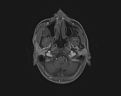
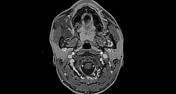
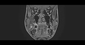
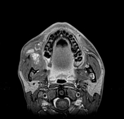
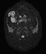
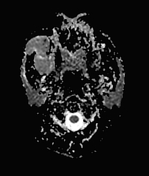
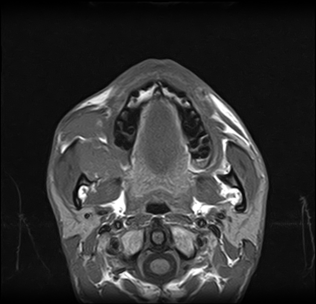
The MRI sequences confirm the location of the lesion within the buccal space, lateral to the buccinator muscle, medial to the masseter muscle, anterior to the medial pterygoid muscle and mandibular ramus, and posterior to the orbicular oris muscles. It appears isointense to the muscles on T1WI, markedly hyperintense onT2WI with foci of calcification (phleboliths). The postcontrast dynamic shows progressive enhancement with partial fill-in on delayed axial sequence (20 minutes).
Case Discussion
Ultrasound and MRI features are most consistent of hemangioma of the buccal space.
Additional contributor/ A. Ramdani, MD


 Unable to process the form. Check for errors and try again.