
Presentation
Known case of a systematic disorder. There is also history of knee trauma. Joint movement is painful and limited.
Patient Data
Age: 7-year-old
Gender: Male
From the case:
Haemophilic arthropathy
Download
Info
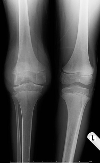
The right knee joint shows moderate effusion, decreased joint space, subchondral erosions, and the physis is narrow compared to the other knee. The intercondylar notch is irregular and wide.
From the case:
Haemophilic arthropathy


Download
Info
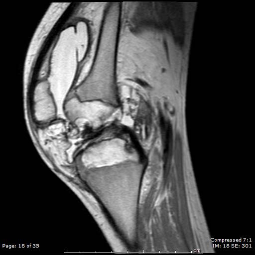
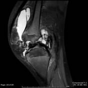
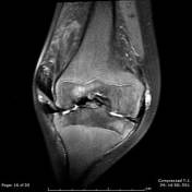
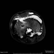
- moderate joint effusion
- decreased joint space
- subchondral erosions and cystic changes
- cartilage loss
- the synovium is thickened with multifocal low signal intensity
- the intercondylar notch is widened with erosive changes
Case Discussion
This young patient has haemophilia A, with multiple previous episodes of knee pain, also he sustained significant knee trauma previously, which altogether lead to these typical findings of advanced or neglected haemophilic arthropathy too early.
Due to repetitive episodes of intra-articular bleeds, the synovium got stained with blood products (haemosiderin), seen as low signal covering the synovium.


 Unable to process the form. Check for errors and try again.