
Presentation
Known case of hypothyroidism with neck swelling associated with dysphagia and dyspnea for four years.
Patient Data

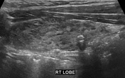
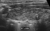
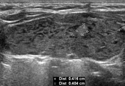
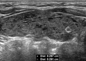
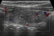
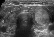
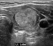
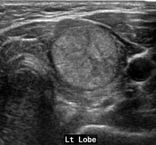
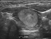
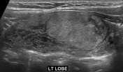
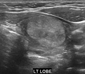
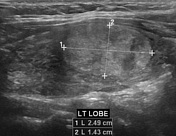
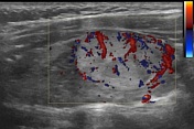
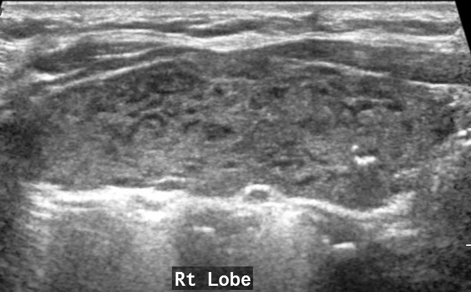
Average size hypoechoic thyroid gland with coarse, heterogeneous parenchymal echotexture and pseudonodular appearance (pseudonodular or giraffe pattern). A hyperechoic nodule measuring 4 x 4 mm and a calcified nodule measuring 3 x 3 mm are seen in the right thyroid lobe. A well-defined hyperechoic nodule measuring 14 x 25 mm is seen in the left thyroid lobe. Increased vascularity is seen in the left thyroid lobe nodule on color Doppler ultrasound examination.

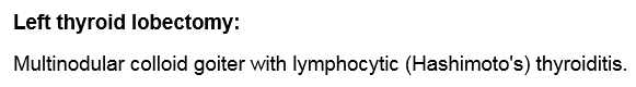
Reports of FNAC (Fine needle aspiration cytology) of the left thyroid nodule and left thyroid lobectomy.
Case Discussion
The patient underwent FNAC of the left thyroid nodule followed by the left thyroid lobectomy and histopathology showed Hashimoto's thyroiditis.


 Unable to process the form. Check for errors and try again.