
Presentation
Reported suspicious mass on ultrasound exam of the liver.
Patient Data
Age: 40 years
Gender: Female
From the case:
Hepatic hemangioma

Download
Info
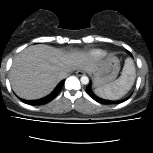
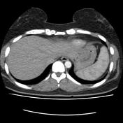
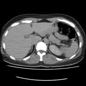
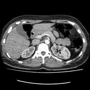
There is a hypodense focus in the right hepatic lobe upper segment VIII subdiaphragmatic with centripetal enhancement characteristic of hemangioma.
Case Discussion
The case is a 40-year-old female with a history of suspicious liver mass lesion on ultrasound exam, triphasic liver MDCT requested, and typical centripetal enhancement of the lesion on the CT images in right hepatic lobe segment VIII subdiaphragmatic region detected. The triphasic MDCT and triphasic MRI of the liver are both useful and standard imaging modalities for liver hemangioma differentiation but MRI has better soft tissue and enhancement resolution.


 Unable to process the form. Check for errors and try again.