
Presentation
Abdominal pain in the right iliac fossa, performing CT scan because of suspected acute appendicitis.
Patient Data
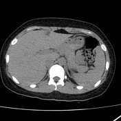

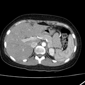
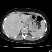
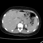

Axial non-contrast, arterial, and portal phase hepatic CT images showed a triangular hypoattenuating lesion in segment IV anteriorly adjacent to the falciform ligament without significant mass effect. A similar lesion was present on the posterior edge of segment VI.
A small appendicolith was seen but the appendix still had a normal diameter of 6 mm, contained intraluminal gas, and was surrounded by homogeneous non-inflamed fat.
A small amount of free fluid was visualized within the pouch of Douglas (considered physiological) because no inflammatory changes was noted in pelvic.
Case Discussion
Hypoattenuating hepatic lesion with the typical triangular shape and location (at the posterior edge of segment IV, around the falciform ligament) is characteristic of pseudolesion near the falciform ligament.
An aberrant vein anastomosing with peripheral portal vein branches (inferior veins of Sappey) has been held responsible for the appearance of this pseudolesion.


 Unable to process the form. Check for errors and try again.