
Presentation
Second episode of seizures during last 2 weeks, no abnormalities in neurological examination.
Patient Data
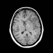

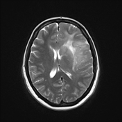
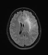
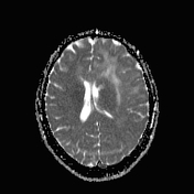

Ill-defined area of increased signal intensity on T2W images affecting left temporal and frontal lobes, with significant mass effect, displacing ventricular system to the right.
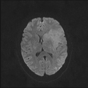
The lesion presents slightly increased signal intensity on DWI (b1000) images. ADC values are similar and slightly higher to the normal white matter.
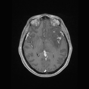
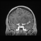
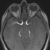
After contrast administration a small focus of enhancement was visualised in left temporal lobe, just adjacent to the MCA. TOF sequences excluded presence of an aneurysm. Contrast administration also revealed presence of a DVA in the left frontal lobe.
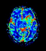
PWI:
Slightly increased rCBV values of peripheral parts of the mass, additionally on the anterior aspect of the lesion there is linear focus of very high rCBV values corresponding to the DVA.
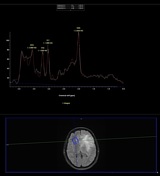
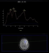
MRS:
The lesion demonstrates pathological brain metabolites spectrum with increased choline-to-creatine ratio and significantly decreased NAA.
Case Discussion
Morphological features of the mass followed by increased rCBV and abnormal spectroscopy suggest a malignant brain tumour. Biopsy was performed and revealed high-grade glioma (WHO grade III).


 Unable to process the form. Check for errors and try again.