
Hyperacute epidural hematoma following ventriculoperitoneal shunt placement
Presentation
Deep coma after 6 hours of wakefulness post VP shunt placement.
Patient Data
8 hrs post-shunt placement
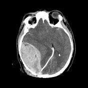

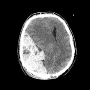
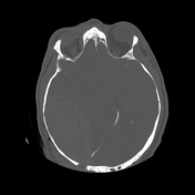

Non-contrast CT shows:
massive Right parietal lentiform well-defined biconvex extra-axial hyperdense collection is noted just anterior to the entry-point of the ventricular catheter, measuring about 12 × 12 × 6 cm, with severe mass effects, midline shift, subfalcine, transtentorial, uncal and tonsillar herniation signs
swirl sign is noted (an isodense to hypodense area represents a hyperacute component indicating unclotted blood/active bleeding inside the hyperdensity that represents an acute component and indicates clotted blood)
the findings correlate with hyperacute epidural hematoma
subfalcine, uncal and cerebellar tonsillar herniation
Case Discussion
EDH is a very rare complication for VP shunt placement, it may occur due to an injury to a meningeal vessel at the entry point of ventricular catheter, or due to blunt insertion of the ventricular catheter without opening the dura, which may allow the dura to be dissected and moved away from the bone, as a result, the epidural space to be created and filled with blood-forming an epidural hematoma.
Swirl sign 1-4 indicates active ongoing bleeding and is associated with poor outcome.
Post-shunting monitoring is so important to detect and manage such an early complication.


 Unable to process the form. Check for errors and try again.