
Presentation
Sudden-onset palatal myoclonus.
Patient Data
Age: 60 years
Gender: Male
From the case:
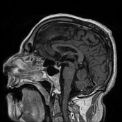
Hypertrophic olivary degeneration


Download
Info
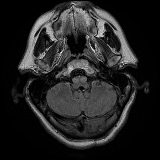
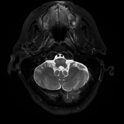
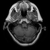
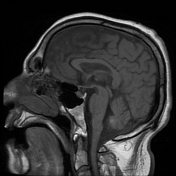
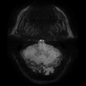
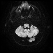
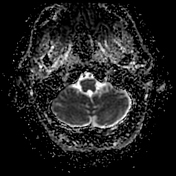
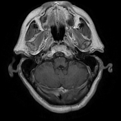
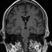
Bilateral symmetric enlargement and mild hyperintensity on T2/FLAIR in the inferior olivary nuclei of the medulla. There is no associated enhancement or susceptibility. No definite lesion is seen in the region of the superior or inferior cerebellar peduncles with a central segmental tract along the pons or in the dentate nuclei.
Case Discussion
This case demonstrates the typical appearance of bilateral hypertrophic olivary degeneration. However, in this case, no lesion was seen in the region of the triangle of Guillain-Mollaret.
It should be noted that a significant number of patients with hypertrophic olivary degeneration will not have an identifiable lesion on MRI 1.


 Unable to process the form. Check for errors and try again.