
Presentation
Severe hepatorenal failure following cardiac arrest. INR 6.
Patient Data
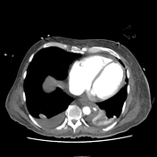


Dilated heart chambers.
Left apical thrombus overlying an area of mural thinning.
Small calibre aorta and branches. Distended IVC and hepatic veins. Contrast medium reflux.
Portal, splenic and SMV thrombosis.
Patchy poor enhancement of the bowel, particularly the distal jejunum and descending colon.
Patchy mucosal hyperenhancement, particularly the duodenum and proximal jejunum.
Oedematous, thick-walled ascending and transverse colon.
Pleural effusions, ascites and generalised oedema including mesentery.
Dilated and displaced oesophagus.
Nodular liver surface.
Hyperenhancing adrenal glands during portal venous phase.
Diminished enhancement in the pancreatic head.
ETT satisfactory.
NGT follows course of dilated oesophagus and indents st
Case Discussion
Alcoholic cirrhosis and dilated cardiomyopathy complicated by myocardial infarction and cardiac arrest. Following resuscitation CT shows LV thrombus, full systemic veins and splanchnic venous thrombosis as well as features of hypoperfusion complex: small arteries and abnormal perfusion of the bowel, adrenal glands and pancreas.


 Unable to process the form. Check for errors and try again.