
Presentation
Persistent cough for many months.
Patient Data
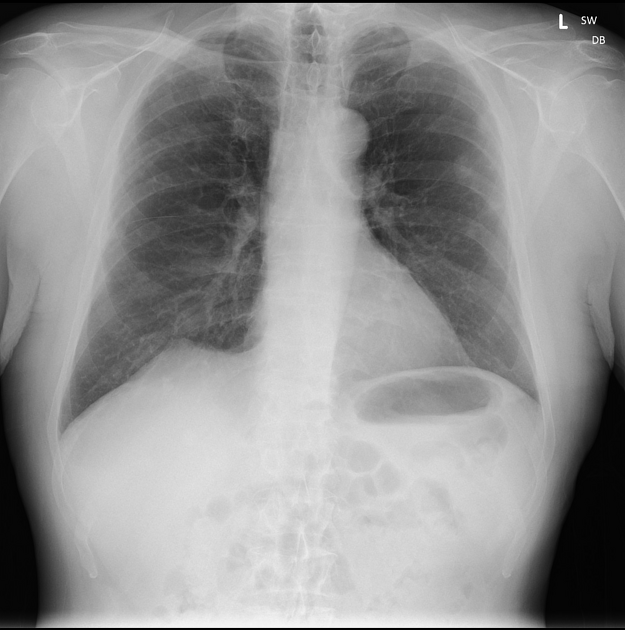
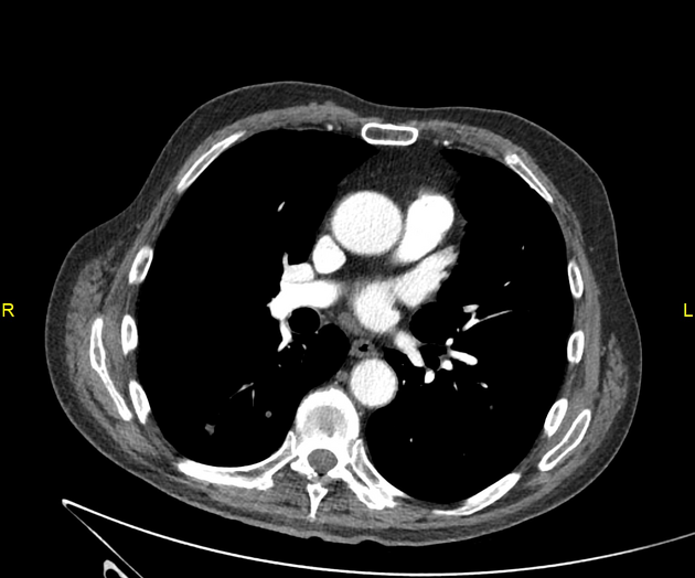
Subtle bilateral lung nodules, likely metastases.
Heart size normal. Normal mediastinal contours.
CXR showed several small opacities projected over the left upper zone, up to 1cm in diameter. Recently reported 2 week history of urinary frequency.
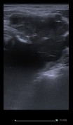
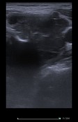
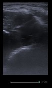
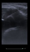
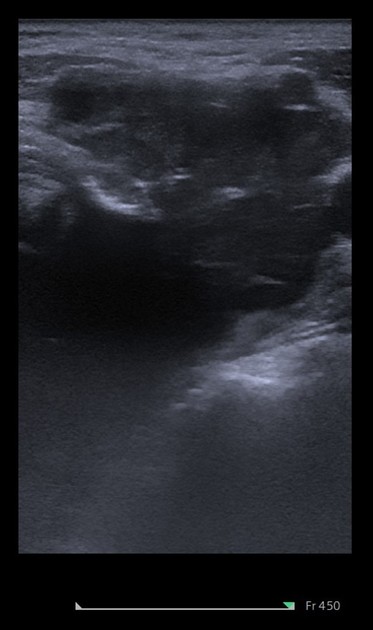
Irregular solid left sided mass at the level of the hypopharynx.
It was amenable to percutaneous ultrasound guided biopsy.


5.5cm left hypopharyngeal mass, partially destroying thyroid and cricoid cartilage and adjacent lymphadenopathy.
No significant mediastinal or axillary lymphadenopathy.
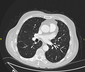
Numerous nodules in both lungs consistent with pulmonary metastasis in the size range of 5-10 mm, largest nodule in the left upper lobe measuring 15 mm.
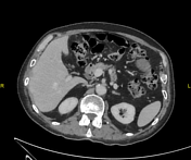
Normal liver, gallbladder, spleen, pancreas, both kidneys and adrenals.
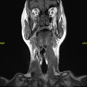
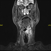
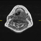
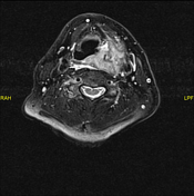
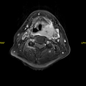
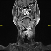
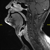
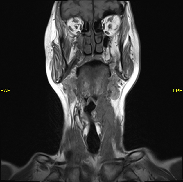
Large tumor in the left side of the neck centered on the left hypopharynx, at the level of the vocal cords. The tumor extends on to towards the pharynx infiltrating across the laryngeal apparatuse, to involve both sides of the thyroid lamina. The tumor extends posteriorly along the prevertebral muscle and it crosses the midline by 1.5 cms. Inferiorly, the tumor extends down into the pyriform fossa where it abuts the left side of the cervical esophagus. Superiorly, it reaches the level of the palatine anteriorly, it extent crossed the posterior half of the thyroid lamina. Laterally, if partially encases the carotid bifurcation.
Several pathological lymph nodes in the left lower neck, the largest measuring 2 cm.
Multiple pulmonary metastases at the lung apices.
Histopathology
Invasive squamous cell carcinoma - head and neck origin.
Case Discussion
Imaging appearances in keeping with a metastatic hypopharyngeal malignancy.
TMN staging : T4b (carotid bifurcation involvement), N3 (pathological nodes below the level of cricoid cartilage). M1.


 Unable to process the form. Check for errors and try again.