
Presentation
Known IIH. New vomiting and disc blurring on optical assessment.
Patient Data
Age: 25 years
Gender: Female
From the case:
Idiopathic intracranial hypertension

Download
Info
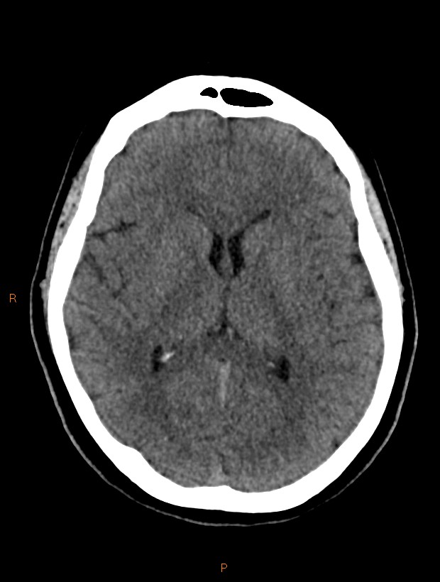
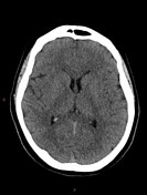
No hematoma or other mass lesion.
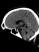
Normal ventricle size, with no critical herniation or tonsillar descent.
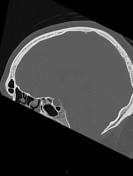
Partly empty pituitary sella / arachnoid herniation, with increased dimension from a study five years earlier.
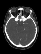
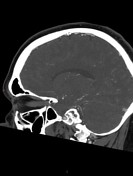
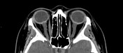
Distention and distortion of the optic nerve sheaths. Protrusion the optic nerve heads.
No venous sinus thrombosis. Some narrowing of the lateral transverse sinuses.
Case Discussion
This case demonstrates some features seen in idiopathic intracranial hypertension, with a particularly good example of radiological papilledema on CT.
The opening pressure on subsequent lumbar puncture was 43 cmH2O.


 Unable to process the form. Check for errors and try again.