
Presentation
Nodules in the scrotal skin for years, patient has cosmetic concerns. Urology service requested ultrasound.
Patient Data

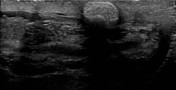
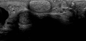
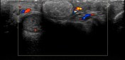
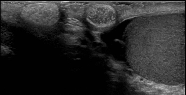
Ultrasound shows oval circumscribed hyperechoic smooth oval avascular nodules in the dermis of scrotum.
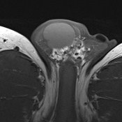

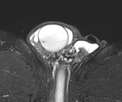
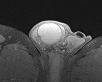
Nodules in the dermis of the scrotum are T1 and T2 hypointense on MRI with no enhancement post contrast suggesting calcified components.
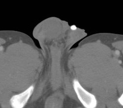
CT scan confirms the calcific nature of these nodules, findings consistent with idiopathic scrotal calcinosis.
Case Discussion
Idiopathic scrotal calcinosis is rare benign condition, characterized by presence of multiple nodules in the dermis of scrotum filled with chalky material/calcium deposits. The diagnosis can easily be confirmed by depiction of calcific content of nodules on imaging.


 Unable to process the form. Check for errors and try again.