
Presentation
Preterm (33 weeks gestation) baby with emergent C-section due to abnormal antenatal scan.
Patient Data


Prominent fluid filled small bowel loops, mild ascites, hydroceles and minimal pleural effusion. Abnormal Doppler ultrasound examination of the umbilical artery (high resistive index with absent diastolic flow). Normal Doppler ultrasound examination of the middle cerebral artery.
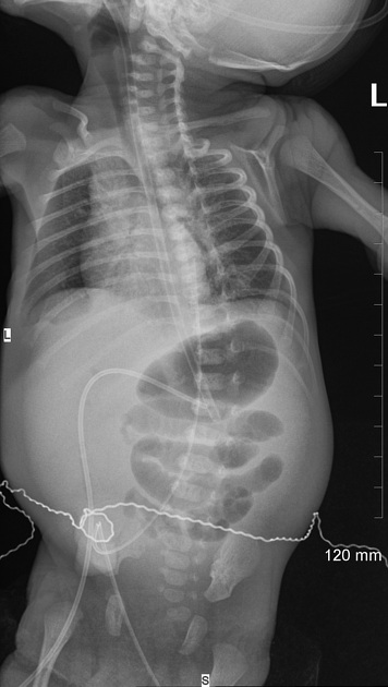
Gas seen in the stomach and proximal small bowel loops. No gas is seen in the distal bowel. No pneumoperitoneum. Appropriately positioned enteric tube. Umbilical catheters are also seen in place; tip of the umbilical arterial catheter (UAC) is projecting over the spine at T8 level whereas the tip of the umbilical venous catheter (UVC) is seen in the epigastric region, likely within the left portal vein which needs adjustment.


Gas filled stomach. Interval development of a radiolucency in the right hypochondrium and epigastrium which is not following the outlines of the stomach or bowel, and is suspicious for pneumoperitoneum (later confirmed by the cross-table and decubitus views). UVC has been withdrawn slightly with its tip now lying in the right hypochondrium (still within the liver/portal vein). Multiple branching radiolucencies, likely representing gas within the portal tributaries, related to the UVC, are seen projecting over the liver shadow.
Case Discussion
the patient underwent an urgent laparotomy
operation notes: Ileal atresia associated with volvulus, 1-2 cm from the ileocecal valve, perforation in the terminal ileum with peritonitis


 Unable to process the form. Check for errors and try again.