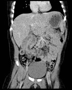
Ileocolic intussusception
Diagnosis certain
Disclosures
- updated 11 May 2022:
Nothing to disclose
Updates to Case Attributes
Body
was changed:
The patient's vague clinical presentation at the outside hospital triggered evaluation with CT rather than the typical workup of ultrasound. Approximately 5 hours later when the patient arrived, ultrasound confirmed continued presence of the intussusception and preserved vascularity. Immediately after ultrasound, the patient was taken for enema reduction, which was successful on the second attempt. (The intussusception easily reduced to the level of the ileocecalileocaecal valve initially but would not easily reflux on the first attempt, as commonly happens).)
-<p>The patient's vague clinical presentation at the outside hospital triggered evaluation with CT rather than the typical workup of ultrasound. Approximately 5 hours later when the patient arrived, ultrasound confirmed continued presence of the intussusception and preserved vascularity. Immediately after ultrasound, the patient was taken for enema reduction, which was successful on the second attempt. (The intussusception easily reduced to the level of the ileocecal valve initially but would not easily reflux on the first attempt, as commonly happens.)</p>- +<p>The patient's vague clinical presentation at the outside hospital triggered evaluation with CT rather than the typical workup of ultrasound. Approximately 5 hours later when the patient arrived, ultrasound confirmed continued presence of the intussusception and preserved vascularity. Immediately after ultrasound, the patient was taken for enema reduction, which was successful on the second attempt. (The intussusception easily reduced to the level of the ileocaecal valve initially but would not easily reflux on the first attempt, as commonly happens).</p>
Updates to Study Attributes
Images Changes:
Image CT (C+ portal venous phase) ( update )
Cropped
image