
Presentation
Post operative case of tracheo-oesophageal fistula. 2D Echocardiography reveals ostium secondum ASD (left to right shunt) with ? double aortic arch.
Patient Data


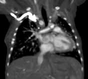
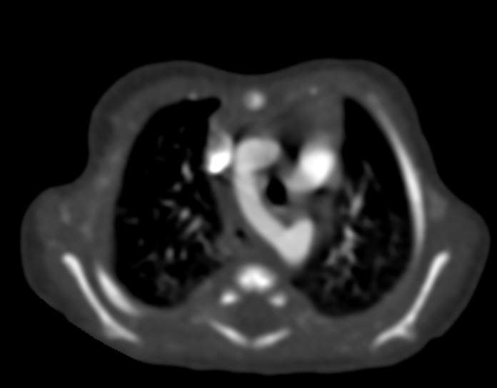
There is evidence of right aortic arch coursing between trachea and oesophagus. The first and second branches arising from right arch are right common carotid and right subclavian artery respectively. Atretic left arch from which left innominate artery is arising which further branches into left common carotid and left subclavian artery.
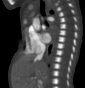
Descending aorta is seen on left side. Small aneurysmal like outpouching measuring approx 4.3mm in diameter and 4.2 mm in length is seen at right aortic arch and descending aorta junction, projecting anteriorly, suggestive of kommerell diverticulum. This diverticulum is residual distal segment of left arch.
Note is made of small residual pouch of treacheo-oesophagal fistula at posterior wall of trachea just before bifurcation.
Small consolidatory changes at right middle lobe.
Imaging features are suggestive of an incomplete double aortic arch and kommerell diverticulum with trachea positioned inside the ring.
Note is also made of almost symmetrical subcutaneous soft tissue enhancement of both upper chest walls, consistent with brown fat enhancement (normally seen on CT angiograms of neonates).
Pectus excavatum is also noted.
Case Discussion
This anomaly results when a segment of the left arch in the embryologic double aortic arch undergoes atresia rather than complete involution between left subclavian artery distal to the left ductus arteriosus and the descending aorta. Now the left innominate artery is seen arising from left arch, followed by the right common carotid and right subclavian artery arising from right arch.
Kommerell diverticulum is remnant of left 4th aortic arch which does not undergo complete involution and in this case, it is basically residual distal atretic segment.
Differential Diagnosis is right aortic arch with mirror image branching which can be ruled out by looking at the position of descending aorta which is positioned on right side in right arch variant and on left side in this case.


 Unable to process the form. Check for errors and try again.