
Presentation
Painful swelling of the right upper neck.
Patient Data

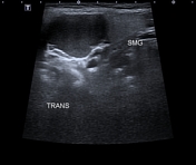

The ultrasound images demonstrate a well-circumscribed ovoid homogeneously hypoechoic cystic lesion with internal debris and posterior acoustic enhancement. This cystic lesion is separated from the adjacent submandibular gland (SMG).


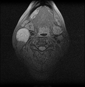
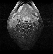
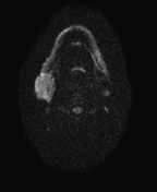
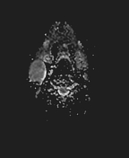
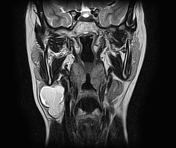
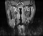
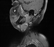
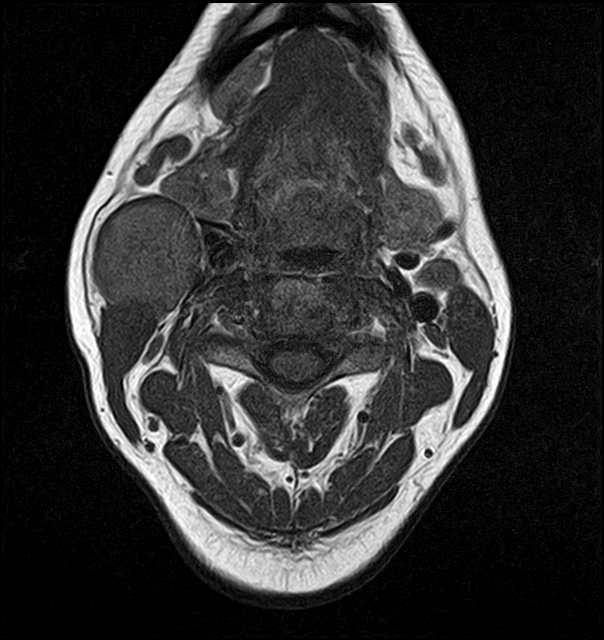
The previously described cystic lesion appears lobulated of slightly high signal on TWI, high signal on T2WI with thick enhancing wall on postcontrast sequences, and restricted diffusion on DWI/ADC. This lesion is located, below the parotid gland, anterior to the sternocleidomastoid muscle (SCM), posterior to the submandibular gland, and lateral to the carotid bifurcation, and internal jugular vein. Small enlarged lymph nodes are noted.
Case Discussion
The ultrasound and MRI features are suggestive of an infected second branchial cleft cyst.
The differential diagnosis should include:
cat-scratch disease
other less common lesions: vascular neoplasms, carotid body tumor, cystic hygroma, ectopic thyroid or salivary tissue, and paraganglioma.


 Unable to process the form. Check for errors and try again.