
Presentation
Left neck pain and swelling.
Patient Data
Age: 20 years
Gender: Male
From the case:
Infected second branchial cleft cyst


Download
Info

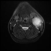
Large cystic lesion at the left submandibular region with the following criteria:
measures 4 x 5 in axial dimensions and 6 cm in CC dimension
low signal on T1, a high signal on T2 and T2 fat suppression sequences
displaces the left submandibular gland anteriorly and sternocleidomastoid muscle posterolaterally
post-contrast sequence reveals thick and irregular wall enhancement of the cyst
Case Discussion
MRI findings are consistent with the infected second branchial cleft cyst.


 Unable to process the form. Check for errors and try again.