
Presentation
Per rectal bleeding.
Patient Data
Age: 60 years
Gender: Male


Download
Info
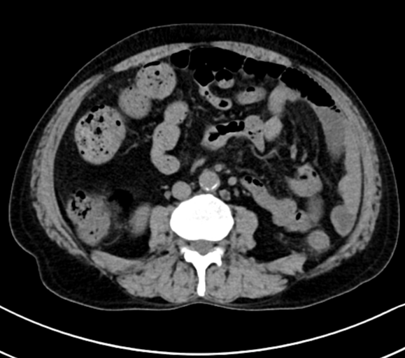
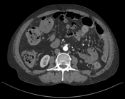
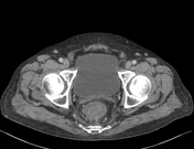
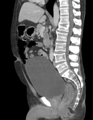
Concentric mural thickening of the rectum, sigmoid colon, descending colon and distal transverse colon with wall stratification and relatively engorged mesenteric vessels.
No lymphadenopathy.
No contrast extravasation.
Case Discussion
CT showed distal colitis and proctitis with submucosal edema. Colonoscopy was performed and the biopsies showed diffuse chronic colitis with moderate activity in keeping with inflammatory bowel disease, favoring ulcerative colitis over Crohn’s disease. No dysplasia.


 Unable to process the form. Check for errors and try again.