
Presentation
The patient presents with multiple pre-syncopal episodes characterised by altered mentation, dizziness, and diaphoresis, which resolved with food. More severe symptoms required urgent transport to the emergency room and inpatient admission. Initial laboratory testing revealed a random blood glucose of 47. Symptoms completely resolved after the administration of a D50 glucose solution. Continued inpatient monitoring revealed recalcitrant hypoglycaemia requiring a D50 drip. After ruling out factitious hypoglycaemia, a multiphase CT of the pancreas was performed.
Patient Data
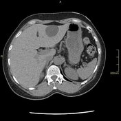

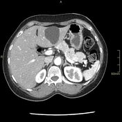
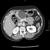
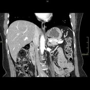
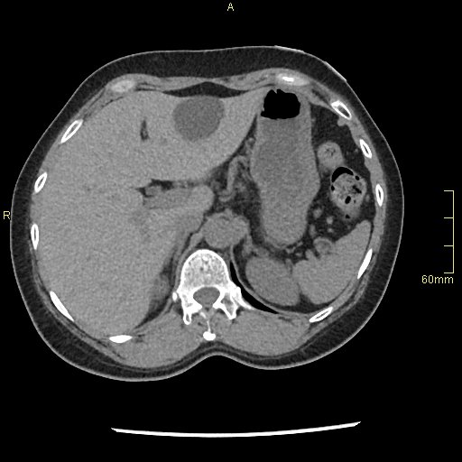
There is a 1.3 cm arterial hyper-enhancing mass within the pancreatic tail. The mass is not visible on non-contrast images and barely perceivable on portal venous phase imaging. The arterial vascular supply of neuroendocrine tumours is a characteristic feature which helps differentiate them from other primary pancreatic neoplasms.
Ga68 DOTATATE PET/CT reveals a pancreatic tail mass with intense somatostatin receptor (SSTR) uptake. This correlates with the mass seen on contrast-enhanced CT.
Case Discussion
This patient presented with repetitive, severe, hypoglycaemic episodes of unknown aetiology. Due to high clinical suspicion of a functional insulinoma, multimodality imaging was utilised to confirm the diagnosis.
Anatomic imaging with an appropriately protocoled multi-phase contrast-enhanced CT demonstrated a well-circumscribed arterial hyper-enhancing mass within the pancreas. Its location and characteristic enhancement pattern strongly suggested a pancreatic neuroendocrine tumour.
Confirmatory molecular imaging with Ga68 DOTATATE PET/CT revealed that the pancreatic mass demonstrated intense somatostatin receptor avidity consistent with a well-differentiated neuroendocrine tumour.
This case beautifully illustrates the use of correlative imaging within the field of molecular imaging. Because of the neuroendocrine tumour's unique combination of anatomic and molecular features, an accurate pre-operative diagnosis can be confidently made.
The mass was resected, and they have returned to a normal state of health.


 Unable to process the form. Check for errors and try again.