
Presentation
Congenital, superficial, swelling adjacent to the right medial canthus.Increasing in size. For operative assessment.
Patient Data


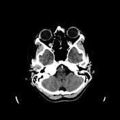

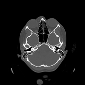
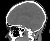
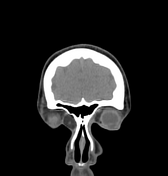
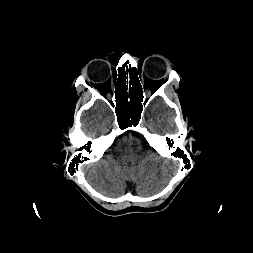
There is a right, superonasal, extrachonal, predominantly fat density, circumscribed, non-enhancing mass. The mass is easily overlooked on the routine pre and post-contrast CT brain windows as demonstrated here. The modified soft tissue reconstructions assist in delineating the mass and appreciating its mixed-density appearance (predominantly fat density -50 Hu, with a central soft tissue nodule). There is minimal underlying bone remodeling. CT imaging is otherwise normal.
Case Discussion
Features consistent with an internal angular orbital dermoid cyst suspected to arise from the frontoethmoidal suture in this instance. The cyst is intact and uncomplicated at the time of imaging.
Case courtesy of Dr S. Palliam.


 Unable to process the form. Check for errors and try again.