
Presentation
Sudden onset headache followed by collapse with right sided weakness.
Patient Data
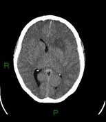

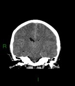
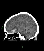

Lobar intracerebral hematoma involving the inferior left frontal lobe. A small volume of perihaematomal edema is present. There is a thin left sided sudural hematoma within the anterior cranial fossa and overlying the left temporal lobe. No intraventricular hemorrhage. Probable tiny volume subarachnoid hemorrhage.
Mass effect with rightward midline shift and effacement of the left lateral ventricle. No hydrocephalus.
No areas of calcification or obvious abnormal vessels related to the hematoma. However, the hematoma is adjacent to the circle of Willis, and an underlying aneurysm needs excluding.
No evidence of small vessel disease.

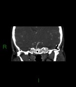
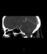
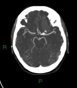
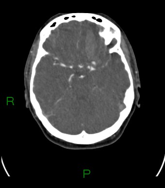
Bi-lobed aneurysm arising from the ophthalmic segment of the left internal carotid artery immediately posterior to the parenchymal hematoma.
Case Discussion
The imaging shows a spontaneous intracerebral hemorrhage secondary to an aneurysm.
- Macrovascular lesions, such as arterial aneurysms, underlie 10-15% of spontaneous intracerebral hemorrhage.
- Early identification of such abnormalities is important to allow appropriate treatment.
- Young age is a risk factor for an underlying macrovascular lesion 1,2, and should lead to vascular imaging.
The patient underwent successful endovascular coiling of the left para-ophthalmic aneurysm.


 Unable to process the form. Check for errors and try again.