
Presentation
Not provided
Patient Data


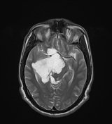
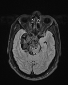
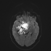
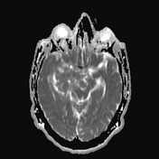
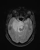
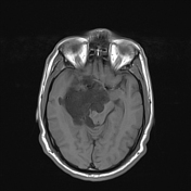
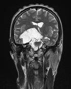
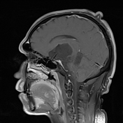
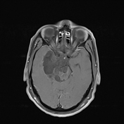
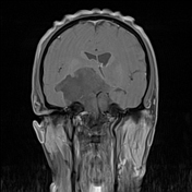
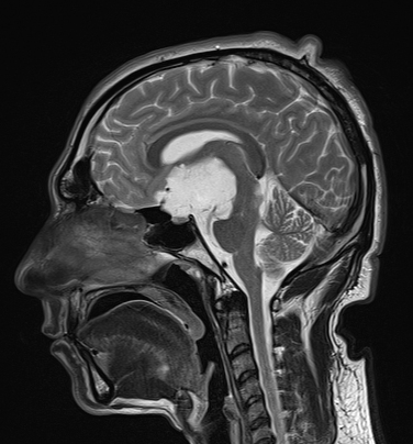
A large well-defined, extra-axial, heterogeneous signal intensity mass lesion extending from the quadrigeminal cistern to prepontine cistern, suprasellar cistern, and medial aspect of right middle cranial fossa. The lesion is returning high signals on T2 weighted images, dirty signals on FLAIR images, low signals on T1 weighted images, and positive diffusion restriction. No significant post-contrast enhancement. No blooming artifact. The lesion abuts the right basal ganglia, the pons, the midbrain, the right thalamus, and the pituitary gland. It is also partially encasing the right middle cerebral artery by the lesion.
There is effacement of the temporal horn of the right lateral ventricle.
No significant hydrocephalus is noted.
No surrounding brain edema is identified.
Case Discussion
The features are suggestive of an intra-cranial large epidermoid cyst.


 Unable to process the form. Check for errors and try again.