
Presentation
Insidious onset of headache (especially when switching to standing position), and intermittent nausea and vomiting.
Patient Data
Age: 25 years
Gender: Female


Download
Info

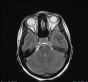
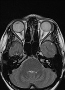
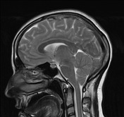
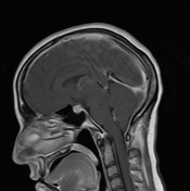
- tonsillar herniation around 6 mm
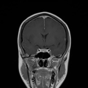
- decreased amount fluid within the optic nerve sheath
- decreased mamillopontine distance (about 1.4 mm)
- decreased pontomesencephalic angle (around 35 degrees)
- mildly enlarged pituitary gland with a superiorly convex roof
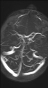
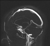
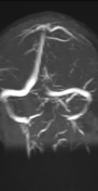
- prominence and rounding (instead of the normal triangular shape in cross-section) of the dural venous sinuses without evidence of thrombosis
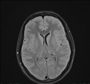
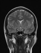
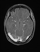
- diffuse pachymeningeal thickening and contrast enhancement
- hypoplastic rostral superior sagittal sinus
From the case:
Intracranial hypotension

Download
Info

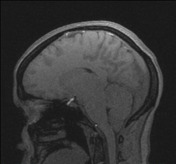
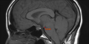
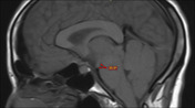
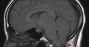
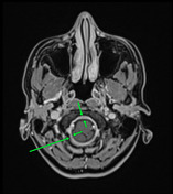
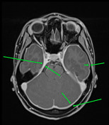
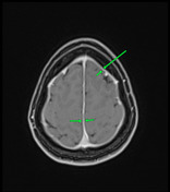
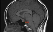
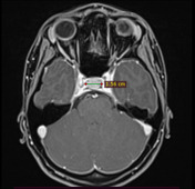
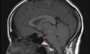
Annotations for some of the above-mentioned findings; the images are arranged in the following order:
- decreased mamillopontine distance (about 1.4 mm).
- decreased pontomesencephalic angle (around 35 degrees).
- tonsillar herniation for around 6mm.
- pachymeningeal thickening and contrast enhancement.
- pachymeningeal thickening and contrast enhancement.
- pachymeningeal thickening and contrast enhancement.
- mildly enlarged pituitary gland with a superiorly convex roof (craniocaudal dimension).
- mildly enlarged pituitary gland (width).
- mildly enlarged pituitary gland (anteroposterior dimension).
- decreased fluid within the optic nerve sheath.
Case Discussion
The clinical features and the imaging appearances are those of intracranial hypotension.


 Unable to process the form. Check for errors and try again.