
Presentation
Late preterm (35/40), male neonate presented with fever, loose motion and poor sucking for last three days.
Patient Data
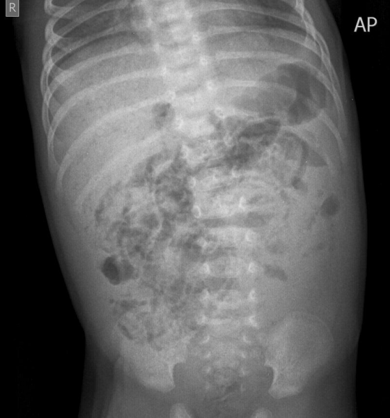
Curvilinear lucencies in bowel walls (intramural gas) and branching lucencies in the liver (portal venous gas).
No abnormal bowel distension or evidence of free intraperitoneal gas.
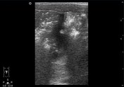

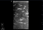
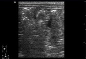
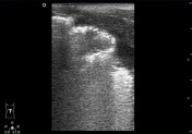
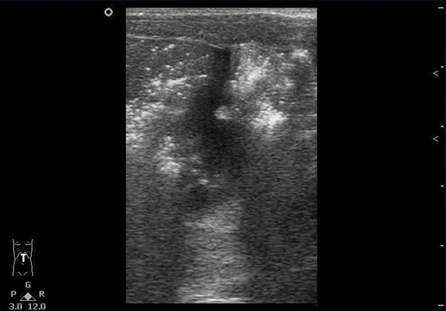
Transabdominal ultrasound with high-frequency linear transducer revealed extensive small echogenic foci in the intestinal walls.
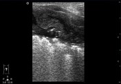
Branching echogenic foci also noted in the liver parenchyma extending to the liver edges representing portal venous gas.
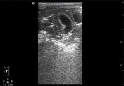
Mild to moderate free fluid was detected in the peritoneal cavity. Mural thickening of the rectum.
Conclusion:
- gas in bowel wall with extraluminal fluid
- rectal mural thickening
- portal venous gas
- ascites
Case Discussion
Intramural gas in the bowel wall is an imaging sign, rather than a diagnosis. This entity can result from various pathologic processes in the neonatal period including prematurity, necrotising enterocolitis, hypoplastic left or right heart syndrome, bowel obstruction, Hirschsprung disease and gastrointestinal rotavirus.
Plain abdominal radiography and ultrasonography are the mainstays of imaging workup. A vertical beam and a horizontal beam radiography with the patient in the supine position are necessary for diagnosis.
Portal venous gas appears as branching structures (lucencies on radiograph or echogenicity on ultrasonography) extending toward the periphery of the liver. The advantage of ultrasound over radiograph is the real time ability to depict bowel mural thickness, echogenicity, peristalsis, mural gas, perfusion and intraperitoneal fluid.
With an appropriate clinical history, the radiologist can play an important role in the detection of pneumatosis intestinalis, differentiation of its medical and surgical causes and finally suggesting an appropriate treatment approach.


 Unable to process the form. Check for errors and try again.