
Presentation
Right proximal-mid third arm anteromedial lump for many years. Gradually increasing in size. Mild local discomfort.
Patient Data


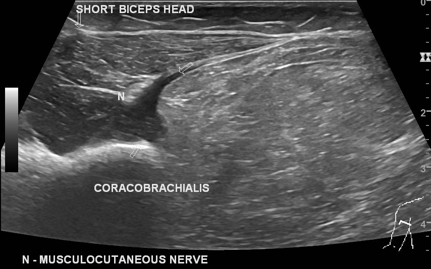
A well-defined solid lesion measuring 120 (craniocaudal) x 70 (transverse) x 46 (anteroposterior) mm. It is located in the medial side of the coracobrachialis muscle, favoured by a claw sign. The long axis of the lesion aligned to the elbow long axis. The cranial end of the lesion is inferior to musculocutaneous nerve origin. The caudal end of the is at the distal end of the coracobrachialis muscle. A patent brachial artery is superficial/ anterior to the lesion. The median and ulnar nerve are along the medial side of the lesion. The musculocutaneous nerve arises cranial to the lesion and its intramuscular segment is lateral and separate from the lesion.
The lesion is hyperechoic to adjacent the subcutaneous fat, and hyperechoic to adjacent muscle with linear incomplete internal striations without calcification/ cystic change/ vascularity. Mildly compressible lesion.
Two short-axis cine-loops run from proximal to distal and cover the entire lesion. 1st loop starts cranial the lesion level. 2nd loops ends after the caudal end of the lesion.
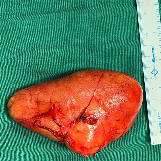
Serial intraoperative photos from the incision, lesion exploration, and excision. Gross specimen photos show a yellowish-coloured encapsulated lesion. The brachial artery is superficial to the lesion.
Intraoperative photos courtesy of operating surgeon Dr Nisarg A. Patel.
Case Discussion
The clinical diagnosis of a long-term large arm lesion in an elderly lady was a lipoma, which was confirmed by ultrasound. The lesion margins were not well-defined on palpation due to its intramuscular location, which becomes more obscured with muscle contraction. This is unlike subcutaneous lipomas, which show distinct palpable margins.
Surgical excision was done. The histopathology suggested a lipoma.


 Unable to process the form. Check for errors and try again.