
Presentation
Headache.
Patient Data
Age: 30 years
Gender: Female


Download
Info
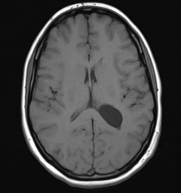
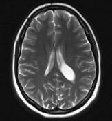
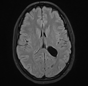
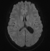
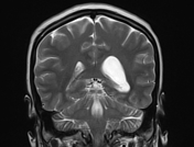
MRI demonstrates a very thin walled ovoid cystic lesion at the posterior part of the body and trigone of the left lateral ventricle which follows CSF on all sequences, including FLAIR and DWI. Slight mass effect, with enlargement of the left lateral ventricular occipital and temporal horns. However, midline is maintained, and no sulcal effacement is present. No MRI findings suspicious for calcification.
Case Discussion
This case demonstrates features consistent with a simple intraventricular cyst. The cyst thin wall is best seen on high-resolution T2 fast spin echo sequences (e.g. CISS) and even on good quality spin echo T2 images.


 Unable to process the form. Check for errors and try again.