
Presentation
The patient has been experiencing ataxia for a few weeks and is being monitored due to HIV infection.
Patient Data
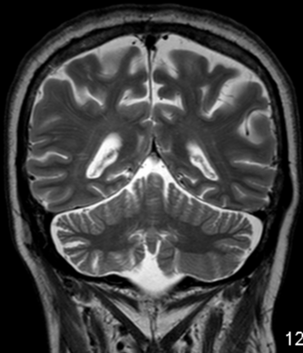
Coronal T2 MRI scan shows subtle atrophy of the right cerebellum without signal intensity abnormality.

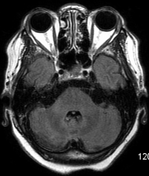
There is worsening of right cerebellar volume loss and subtle areas of high signal in right MCP on axial FLAIR.


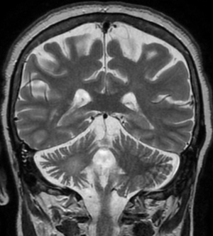
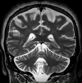
There is a deterioration in the right cerebellar atrophy and a FLAIR hyperintensity is observed in the middle cerebellar peduncles.
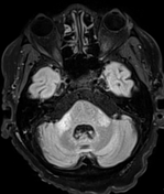
Despite undetectable viral load, radiologic findings have worsened:
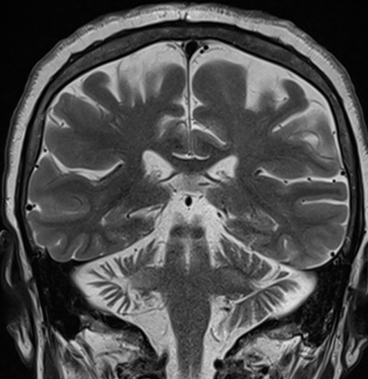
there is bilateral cerebellar atrophy, particularly on the right accompanied by a high T2 signal in both MCP and in the right cerebellar hemisphere
there is no significant supratentorial atrophy
Case Discussion
Clinical presentation, history of HIV infection, cerebellar symptoms, laboratory tests (PCR JC Virus), and radiological findings are nearly pathognomonic of JC virus granule cell neuronopathy.
Granule cell neuronopathy (GCN) is a rare form of CNS JC virus disease first described by Du Pasquier et al. in 2003 and later by Koralnik et al. in 2005. It mainly occurs in immunocompromised patients and is characterised by selective infection of the cerebellar granule cell layer. Patients with GCN typically present with cerebellar symptoms, while imaging studies show cerebellar atrophy and/or signal changes.


 Unable to process the form. Check for errors and try again.