
Presentation
Road traffic accident. Motorcycle versus car.
Patient Data
Radiopaedia's Trauma Radiology Course - Video On-Demand
PEG view demonstrates lateral displacement of the lateral masses of C1 with respect to C2 meaning the bony ring of C1 must be disrupted (normally the lateral bony margins of C1 should not overhang C2). Lateral views of the cervical spine (with a hard collar applied) demonstrate widening of the atlantodens interval and a lucency (fracture) can also be seen traversing the posterior arch.
CT through C1 confirms the plain film findings. Two fractures are identified: on the right through the lateral aspect of the posterior arch; on the left through the lateral aspect of the anterior arch. A further fracture fragment is noted posterior to the dens (which appears normal), representing an avulsion fracture of the transverse atlantal ligament from the right lateral mass of C1.
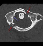
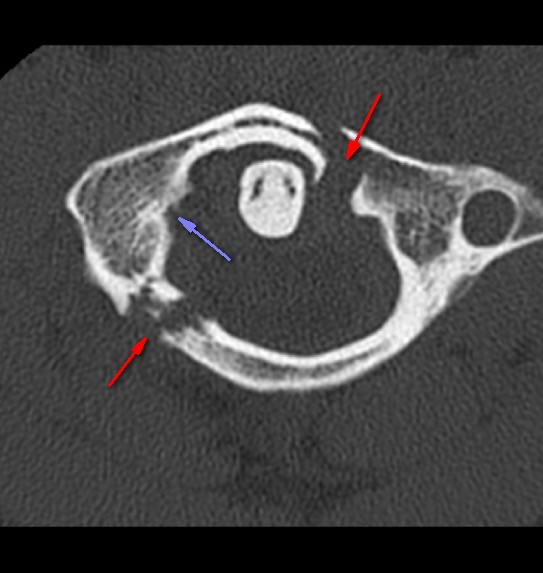
Fractures (red arrows) and avulsion donor site at attachment of atlantal transverse ligament (purple arrow).
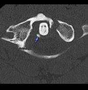
Avulsion fragment (purple arrow) attached to transverse atlantal ligament.
Case Discussion
Jefferson fractures typically occur as a result of axial loading of the head. They can be treated conservatively provided the transverse atlantal ligament is intact, and as such careful examination of the atlantodens interval and presence of an avulsion fracture is critical.
This injury is a Gehweiler type 3b injury, subtype 2.


 Unable to process the form. Check for errors and try again.