
Presentation
Fall term female baby (40 weeks) with a positive family history of previous two siblings with Joubert’s syndrome-related disorder, for MRI brain evaluation.
Patient Data


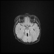
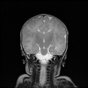

- hypoplasia of the cerebellar vermis with thinning and elongation of the superior cerebellar peduncles giving rise of molar tooth appearance and batwing configuration of the 4th ventricle
- cavum septum pellucidum is noted (normal variant)
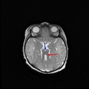

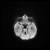
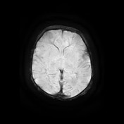
Axial T2-weighted MR image shows the classic malar tooth sign, including thickened, elongated, parallel, and horizontally orientated superior cerebellar peduncles (red arrow) and a deepened interpeduncular fossa (blue arrow).
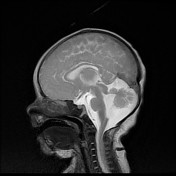
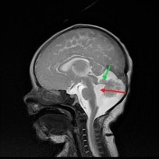
Midsagittal T2-weighted MR image demonstrates a severe vermian hypoplasia-dysplasia (green arrow), and distortion and enlargement of the fourth ventricle (red arrow), and a narrow pontomesencephalic isthmus.
Case Discussion
MRI brain findings are consistent with Joubert syndrome and when associated with anomalies of the kidneys as our case a renal US (not provided ) show left multicystic dysplastic kidney, suggesting of Joubert syndrome-related disorders.


 Unable to process the form. Check for errors and try again.