
Presentation
Term baby (40 weeks) with a positive family history of previous 4 siblings diagnosed with Joubert’s syndrome, for MRI brain evaluation.
Patient Data
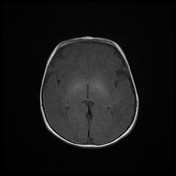


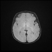
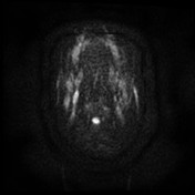
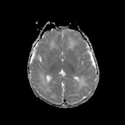
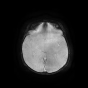

Axial sequences including (T1, T2, FLAIR, DWI and SWAN) weighted MR image showing the classic molar tooth sign, including thickened, elongated, parallel, and horizontally orientated superior cerebellar peduncles and a deepened interpeduncular fossa.
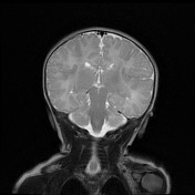
Midsagittal T2-weighted MR image demonstrates a severe vermian hypoplasia-dysplasia and distortion and enlargement of the fourth ventricle and a narrow pontomesencephalic isthmus.
Cavum septum pellucidum is noted (normal variant).
MRI study findings are consistent with Joubert syndrome
Case Discussion
Joubert syndrome is a rare autosomal recessive disorder characterised by neuropathologic abnormalities of the cerebellum and brainstem including inherited hypoplasia or aplasia of the cerebellar vermis with thickening and elongation of the superior cerebellar peduncles giving rise to molar tooth appearance and bat-wing configuration of the 4th ventricle.


 Unable to process the form. Check for errors and try again.