
Presentation
Gobal developmental delay and hypotonia.
Patient Data
Age: 4 years
Gender: Male
From the case:
Joubert syndrome


Show annotations
Download
Info
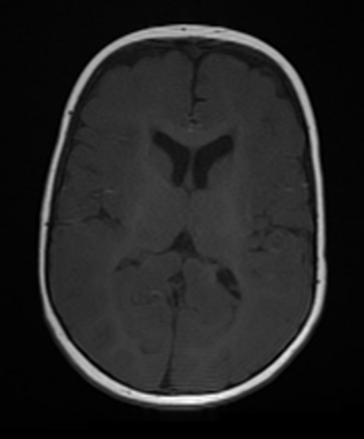
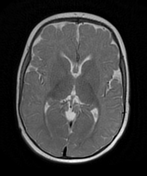
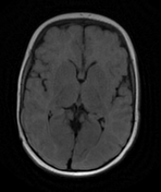
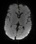
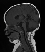
normal size and configuration of the 3rd and lateral ventricles
no midline shift
abnormal configuration (batwing) high riding 4th ventricle
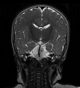
enlarged elongated superior cerebellar peduncles with molar tooth sign
absence of the cerebellar vermis with apposition of both cerebellar hemispheres abutting each other without fusion forming a median cleft
dysplastic both cerebellar hemispheres
no definite intra or extra-axial areas of abnormal intensity
Case Discussion
This case illustrates the typical features of Joubert syndrome, with the superior cerebellar peduncles displaying the appearance of a molar tooth and the 4th ventricle displaying a batwing configuration.


 Unable to process the form. Check for errors and try again.