
Presentation
Known case of joubert syndrome with hypertension and end-stage renal disease.
Patient Data


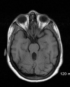
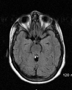
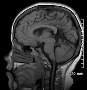
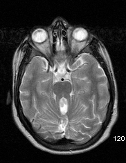
Elongated superior cerebellar peduncles giving the midbrain a classical molar tooth shape, cerebellar vermis hypoplasia and umbrella or bat wing configuration of the 4th ventricle.

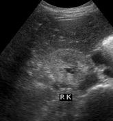
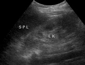
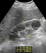
Right kidney measures 10.3 cm and left kidney measures 10.5 cm in length. Both kidneys show increased renal cortical echogenicity with reduced corticomedullary differentiation. No calculi, cysts or hydronephrosis is seen. These findings are consistent with chronic kidney disease.
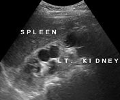
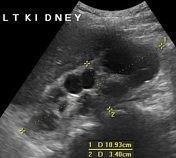
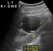
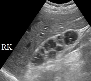
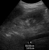
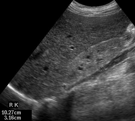
Re-demonstration of increased renal cortical echogenicity bilaterally with interval development of decreased cortical thickness and multiple cysts; the largest cyst seen at the lower pole of the left kidney measures 3.6 cm in diameter. These findings are consistent with acquired cystic kidney disease (patient is a known case of end-stage renal disease and is on hemodialysis for the last three years).
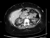
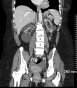
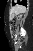
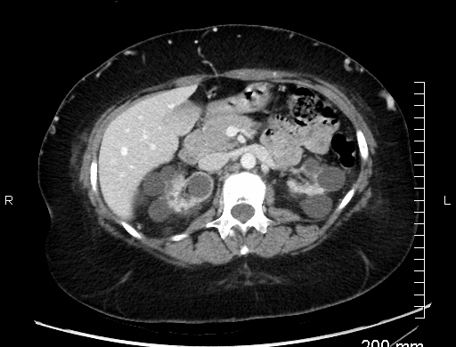
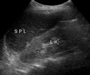
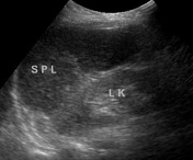
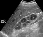
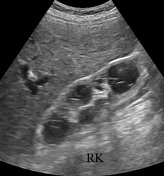
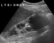
Right femoral permacath is seen in place with its tip lying in the IVC below the renal veins. No signs of IVC obstruction noted. Innumerable variable size simple cysts in both kidneys, in keeping with acquired cystic kidney disease (in this patient with history of long-standing hemodialysis). Multiple venous collaterals noted along anterior abdominal wall (history of chronic superior vena cava obstruction secondary to the dialysis catheters). Simple left adnexal cyst measuring 3.5 x 4.2 cm.
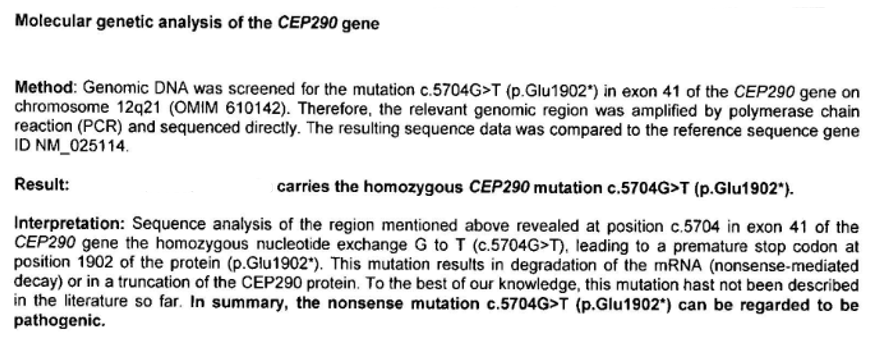
Genetic analysis report is positive for homozygous mutation responsible for the Joubert syndrome.
Case Discussion
Brain imaging findings and genetic analysis are consistent with the diagnosis of Joubert syndrome. Renal imaging findings and history of hemodialysis are consistent with end-stage renal disease (ESRD) with development of acquired cystic kidney disease on the follow-up imaging. Combination of these brain and renal imaging findings is consistent with Joubert syndrome related disorders (JSRD).


 Unable to process the form. Check for errors and try again.