
Presentation
Previous history of melanoma, erosive petrous bone lesion found on CT brain (not shown).
Patient Data


At the left petrous apex region, expanding the jugular foramen and extending into the left middle ear cavity, there is a vividly enhancing solid mass with elevated T2 signal and scattered flow-voids. The lesion has a subcentimetre protrusion abutting the cerebellum in the cerebellopontine angle. Partial obliteration of the mastoid air cells on the left.
Preserved left ICA flow-voids. Dural venous sinuses are patent, with normal opacification of the left sigmoid sinus and favourable torcular anatomy with good communication between both transverse sinuses.
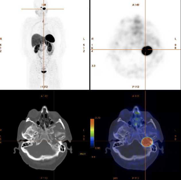
Left petrous bone lesion is intensively octreotate avid, which is best explained by a jugulotympanic paraganglioma given the MRI appearances.
Case Discussion
Although the imaging features on MRI were quite suggestive of a paraganglioma, given the history of melanoma, functional imaging with a Gallium-68 DOTATATE PET CT was performed.


 Unable to process the form. Check for errors and try again.