
Presentation
Epigastric pain, abdominal distension, and looking ill. Ultrasound reported bilateral adnexal masses.
Patient Data
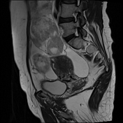

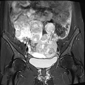
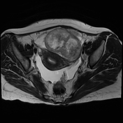
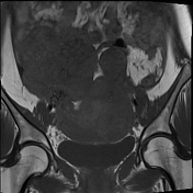
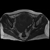
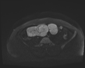
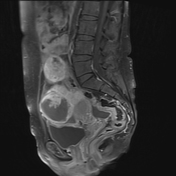
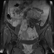


Large predominantly solid masses with necrotic areas in bilateral adnexal regions extending to the right iliac fossa.
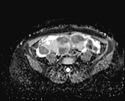
Mild to moderate pelvis ascites.
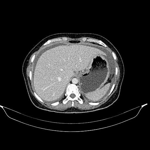
There is a marked circumferential mural thickening of the gastric body and antrum, with a unilateral wall thickness of about 3.3 cm.
Multiple enlarged necrotic lymph nodes are noted posterior to the gastric cardia. The largest measures 3.2 cm in short diameter.
Moderate ascites with some omental thickening.
There are large, solid lesions with necrotic changes in the bilateral adnexal region.
Bilaterally, mild hydronephrosis and hydroureter are likely due to pelvic masses.
Case Discussion
The features suggest gastric primary neoplastic growth with enlarged necrotic lymph nodes posterior to the gastric cardia, ascites, and some omental thickening (peritoneal carcinomatosis).
Bilateral adnexal lobulated masses, in keeping with the gastric neoplastic process, are likely to be adnexal metastasis (Krukenberg tumor) rather than primary adnexal malignancy.


 Unable to process the form. Check for errors and try again.