
Presentation
Painful swelling of lower extremity, walking with limp.
Patient Data

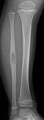
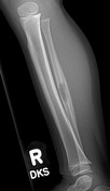

Lytic lesion with somewhat permeative appearance centred in the medullary canal of the midshaft fibula. The lesions features ill defined margins (wide zone of transition). Periostitis surrounding the bone appears in a lamellated pattern, which nearly give the fibular diaphysis an expansile appearance. Extensive soft tissue swelling associated with this lesion.
Histopathology
Microscopic description
The provided slides show aggregates of histiocytes mixed with abundant eosinophils and lymphocytes. Nuclear grooves are also seen in some of the histiocytes. The provided immunostains show that these clusters of histiocytes are positive for CD1a. The morphology and immunoprofile support the diagnosis of Langerhans cell histiocytosis.
Diagnosis
Langerhans cell histiocytosis
Case Discussion
Despite its somewhat atypical location in the fibula, the radiographic appearance and demographic are classic for Langerhans cell histiocytosis.
There are multiple features which connote that this is an aggressive lesion:
- wide zone of transition
- lamellated pattern of periostitis - suggests that physiologic process of bone healing cannot "keep up" with the tissue destruction encited by the lesion
- associated soft tissue swelling
In the absence of signs or symptoms of infection to suggest osteomyelitis, the differential for an aggressive-appearing lesion in a child includes metastasis (e.g. neuroblastoma), blue round cell tumour, and primary osseous lymphoma.


 Unable to process the form. Check for errors and try again.