
Large cerebellopontine angle meningioma with obstructive hydrocephalus
Diagnosis probable
Disclosures
- updated 11 May 2022:
Nothing to disclose
Updates to Study Attributes
Findings
was changed:
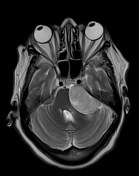
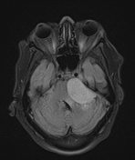
Large extra-axial dome-shaped dural-based mass lesion at left cerebellopontine angle with dural tail. It shows homogeneous texture with smooth outlinesurfaces, and no cystic changes. It is of isointense signal on T1, high signal on T2 and FLAIR.
It is resting on left internal acoustic meatus with minimal intracanalicular extension. Normal diameter of left intermal auditory canal with visualised intracanalicular VII and VIII nerves. ItAnteriorly, it shows extension to the left Meckel cave. Inferiorly it is encroaching upon left hypoglossal canaljugular foramen opening.
It is compressing the brainstem from left side and left cerebellar hemisphere causing moderate supratentorial hydrocephalus with mild periventricular edema.
Images Changes:
Image MRI (T2) ( update )
Cropped
image
Image MRI (FLAIR) ( update )
Cropped
image
Updates to Case Attributes
Title
was changed:
Large cerebellopontine angle meningioma with obstructive hydrocephalus
Diagnostic Certainty
was set to
.
Visibility
changed from unlisted to public.
Updates to Link Attributes
Title
was removed:
Type
was removed.
Visible
was set to
.


 Unable to process the form. Check for errors and try again.