
Presentation
Dysphagia.
Patient Data


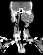
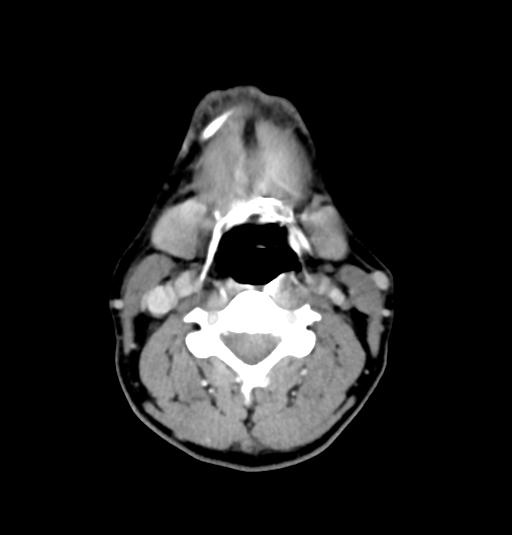
Large heterogeneously enhancing mass involving both the superficial and deep portions of the left parotid gland. The left parapharyngeal space is effaced and medially displaced. Nasopharyngeal airway is narrowed. Carotid space is displaced posteriorly.
Left internal jugular vein is compressed. No suspicious lymph node.
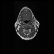
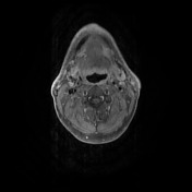
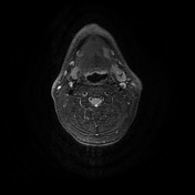
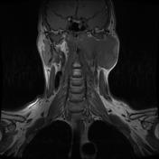
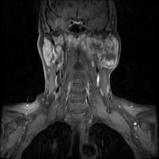
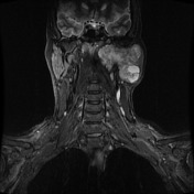
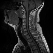

Large mass involving both the superficial and deep portions of the left parotid gland. It has low T1 signal, high but heterogeneous T2 signal, and also demonstrates heterogeneous enhancement. Left parapharyngeal space is effaced and medially displaced.
The patient underwent resection of this mass.
Histopathology
MACROSCOPIC DESCRIPTION: "Left parapharyngeal mass": 88g, 75x60x35 mm. The tissue is white, firm, nodular and with a small piece of skeletal muscle attached at one aspect 50x30x14 mm. The specimen is inked green. The specimen is unoriented. On cut section the specimen has a white and pale yellow variegated cut surface, and there is a central cavity measuring up to 21mm in maximum dimension that is irregular in shape and contains thin brown fluid. The mass is circumscribed but does not have a capsule and extends all the way to the green ink.
MICROSCOPIC DESCRIPTION: The sections show an irregularly shaped tumour with a thin, mostly complete capsule. The tumour forms serpiginous shaped areas, tubules and strands of epithelial and myoepithelial cells. The cells are set within a hypocellular myxocollagenous and cartilaginous matrix. There are no mitoses or necrosis. In the areas where the capsule is present there is no capsular breach. At one of the points where the capsule is missing the tumour is present at the green inked margin. There is no lymphovascular invasion or perineural infiltration. The tumour measures 75 mm in maximum dimension. There is no evidence of malignancy.
DIAGNOSIS: Pleomorphic adenoma.
Case Discussion
Knowing the deep spaces of the head and neck is important to first localise where the mass is, before coming up with a differential diagnosis as this differs greatly from space-to-to-space.


 Unable to process the form. Check for errors and try again.