Large pleomorphic adenoma
Diagnosis certain
Updates to Case Attributes
Diagnostic Certainty
was set to
.
Age
was set to
40.
Gender
was set to
Female.
Presentation
was added:
Chewing and swallowing difficulty, with left TMJ region pain.
Body
was changed:
Large pleomorphic adenoma.
Histology
Left parapharyngeal mass, resection: Pleomorphic adenoma.
Gross description
Received is a single container consists of a rubbery nodule weighing 26 g and measuring 5 x 4.5 x 2.7 cm. Smooth external surface is inked in black. A cross-section of the nodule shows fibrous homogeneous structure with a possible central area of scarring.
-<p>Large <a href="/articles/pleomorphic_adenoma" title="Pleomorphic adenoma">pleomorphic adenoma</a>.</p><h4>Histology</h4><p>Left parapharyngeal mass, resection: Pleomorphic adenoma.</p><h5>Gross description</h5><p>Received is a single container consists of a rubbery nodule weighing 26 g and measuring 5 x 4.5 x 2.7 cm. Smooth external surface is inked in black. A cross-section of the nodule shows fibrous homogeneous structure with a possible central area of scarring. </p>- +<p>Large <a href="/articles/pleomorphic-adenoma-of-the-salivary-glands">pleomorphic adenoma</a>.</p><p>Histology</p><p>Left parapharyngeal mass, resection: Pleomorphic adenoma.</p><p>Gross description</p><p>Received is a single container consists of a rubbery nodule weighing 26 g and measuring 5 x 4.5 x 2.7 cm. Smooth external surface is inked in black. A cross-section of the nodule shows fibrous homogeneous structure with a possible central area of scarring.</p>
Updates to Study Attributes
Findings
was added:
A large left parapharyngeal heterogenously mild enhancing solid mass lesion with some calcifications, reaching close to pterygoid muscles, and causing oropharyngeal attenuation.
Images Changes:
Image CT (C+ arterial phase) ( update )
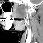
Perspective
was set to
Sagittal.
Specifics
was set to
C+ arterial phase.
Image CT (C+ arterial phase) ( update )

Perspective
was set to
Coronal.
Specifics
was set to
C+ arterial phase.
Image CT (C+ arterial phase) ( update )

Perspective
was set to
Axial.
Specifics
was set to
C+ arterial phase.