
Presentation
Large left pectoral swelling, on heparin for DVT. IVDU.
Patient Data


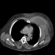
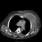
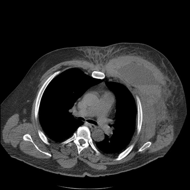
Large left subpectoral hematoma extending superiorly from the left clavipectoral fascia to 5th rib inferiorly and is bounded medially by the sternum and laterally by the axilla. There is evidence of active arterial bleeding but no large artery in continuity with the blushes is identified. Hyperdense layering at the fluid-fluid level seen on the delayed imaging consistent with blood pooling.
Normal opacification of the left subclavian artery. Left internal mammary artery is distant to the collection and patent. Superior thoracic artery passes anterior to the collection to supply the pectoral muscles. No continuity between the axillary artery branches and the arterial blushes identified. Imaged proximal axillary artery is patent. No underlying acute rib fractures.
Right internal jugular line central venous catheter is well positioned.
Nodular air space opacification within the right middle lobe and a cavitating lesion in the posterobasal left lower lobe. No pneumothorax or pleural effusion.
Case Discussion
As there was no bleeding source identified and the patient was stable, DSA was not performed. The patient was treated with US-guided percutaneous drainage of the hematoma and recovered well.
Antibiotic therapy successfully treated the pulmonary disease.


 Unable to process the form. Check for errors and try again.