Left carotid body paraganglioma
Updates to Case Attributes
The differential diagnosis is quite limited in this case, as there is a classic salt & pepper appearance of the lesion on T1 weighted-images images and splaying of the ICA / ECA. This is almost pathognomonic of carotid body paraganglioma.
The differential diagnosis for a tumortumour in this location includes :
- vagal schwannoma: tends to displace both vessels together rather than splaying them
- vagal neurofibroma: tends to displace both vessels together rather than splaying them
- lymph node mass: may look similar if
hyper-vascularhypervascular - glomus vagale tumour: same pathology but located more rostrally
- carotid bulb ectasia
-<p>The differential diagnosis is quite limited in this case, as there is a classic salt & pepper appearance of the lesion on T1 weighted-images and splaying of the ICA / ECA. This is almost pathognomonic of carotid body paraganglioma.</p><p>The differential diagnosis for a tumor in this location includes :</p><ul>- +<p>The differential diagnosis is quite limited in this case, as there is a classic salt & pepper appearance of the lesion on T1 weighted images and splaying of the ICA / ECA. This is almost pathognomonic of carotid body paraganglioma.</p><p>The differential diagnosis for a tumour in this location includes :</p><ul>
- +<li>vagal schwannoma: tends to displace both vessels together rather than splaying them</li>
- +<li>vagal neurofibroma: tends to displace both vessels together rather than splaying them</li>
- +<li>lymph node mass: may look similar if hypervascular</li>
-vagal schwannoma : tends to displace both vessels together rather than splaying them</li>-<li>-vagal neurofibroma : tends to displace both vessels together rather than splaying them</li>-<li>lymph node mass : may look similar if hyper-vascular</li>-<li>-<a href="/articles/glomus-vagale-tumour-3">glomus vagale tumour </a>: same pathology but located more rostrally</li>- +<a href="/articles/glomus-vagale-tumour-3">glomus vagale tumour</a>: same pathology but located more rostrally</li>
Systems changed:
- Oncology
- Head & Neck
Updates to Study Attributes
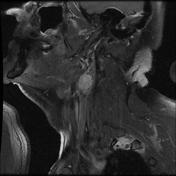
MRI shows a round lesion located between the internal and external carotid bifurcation, iso-intense to muscle on T1 & T2 (salt & pepper apperanceappearance) & T2, and enhancing avidly after gadolinium administration. The internal and external carotid arteries are splayed by the lesion.
The most probablyprobable diagnosis is a carotid body paranganglioma (or carotidparaganglioma (carotid glomus tumor, / chemodectoma, )
Image MRI (T1) ( update )
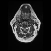
Image MRI (T1 fat sat) ( update )
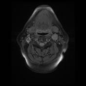
Image MRI (T2) ( update )
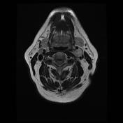
Image MRI (T2) ( update )
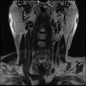
Image MRI (T1 C+ fat sat) ( update )
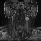
Image MRI (T1 C+ fat sat) ( update )
Updates to Study Attributes
MRI TOF revealed the enhancing left carotid bifurcation lesion, splaying each of carotid artery branches.
Image MRI (TOF) ( update )
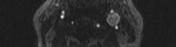
Image MRI (TOF) ( update )
