
Presentation
The patient presented to the emergency department with abdominal pain and hypotension.
Patient Data
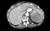


There is a moderate amount of acute hemoperitoneum secondary to active bleeding in the gastrohepatic region due to an arterial pseudoaneurysm vs ruptured aneurysm arising from the left gastric artery.
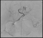
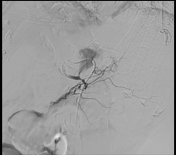
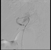
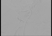

There is a left gastric artery pseudoaneurysm vs ruptured aneurysm with a narrow short neck. Subsequent successful embolization of the left gastric artery distal and proximal to the level of the pedunculated pseudoaneurysm was performed. Closure of the front door and back-door of the pseudoaneurysm was achieved with no residual filling of the lesion on the later images.
Case Discussion
Our patient presented to the emergency department with abdominal pain and syncope. Urgent CT was done and revealed hemoperitoneum secondary to active bleeding. A left gastric artery pseudoaneurysm/ruptured aneurysm was identified and successfully repaired with coil embolization.
Characteristic imaging findings of a gastric artery aneurysm include arterial ballooning with or without hemoperitoneum due to bleeding.
Case co-author: Ashwin Hampole, MD, Advocate Good Samaritan Hospital, USA.


 Unable to process the form. Check for errors and try again.