Presentation
Cough and dyspnea, on physical examination decreased air entry on the left side.
Patient Data
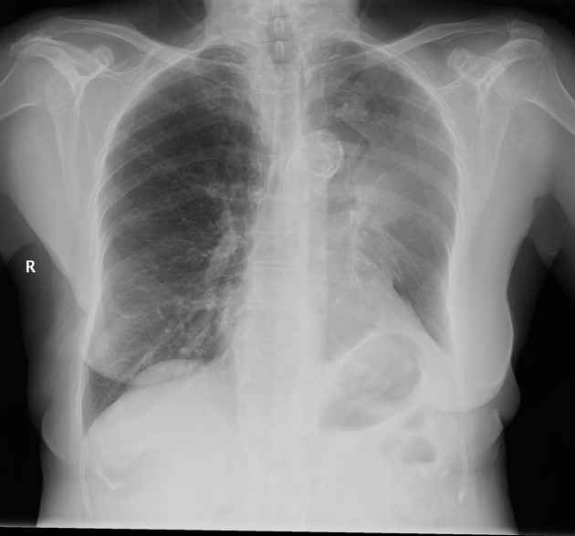
CHEST X- RAY:
Hazy (or veil-like) and decreased volume of the left hemithorax are seen.
Lucency adjacent to the aortic knuckle is seen mostly due to collapsed left upper lobe (luftsichel sign).
Left hilar opacity of soft tissue density is seen.
Tenting of the left hemidiaphragm.
Clear right lung and right costophrenic angle.
Chest CT scan with IV contrast is advised for better evaluation.
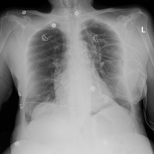
CHEST X-RAY:
Compared to previous chest x-ray done two days ago, more clearance of the left hemithorax is noted with re-expanded left lung.
Blunted left costophrenic angle

CHEST X-RAY:
Compared to previous chest x-ray done two days ago.
Left hilar enlargement is seen due to the known mass lesion.
Emphysematous lung changes are seen.
Normal cardio thoracic ratio and atherosclerotic aorta is noted.
Clear costophrenic angles.
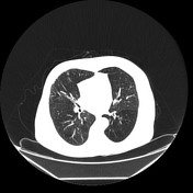

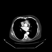

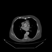

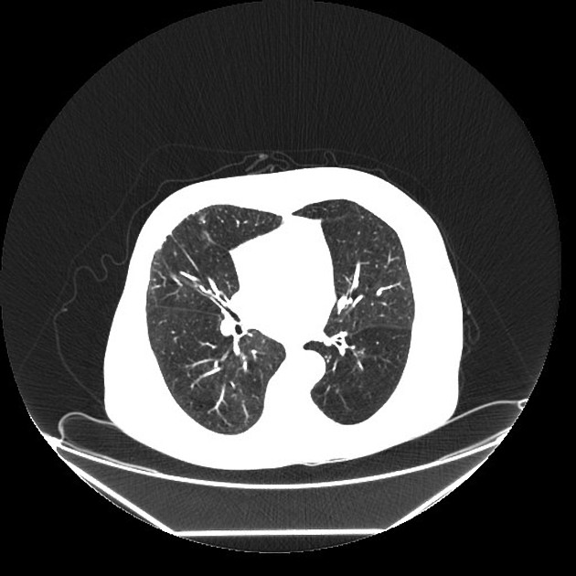
CHEST CT SCAN WITH CONTRAST ENHANCEMENT:
There is a spiculated solid mass about 2.2 cm in diameter in the left hilum consistent most likely with central bronchogenic carcinoma.
Two enlarged lymph nodes are seen at the aorta-oulmonary window the largest measures 2.5 cm in its long axis.
Fine nodular pleural thickening is seen in the right side superiorly and anteriorly, so pleural metastases could not be excluded.
No definite evidence of pulmonary metastases.
No evidence of pleural effusion.
No evidence of bony metastases.
Dense spiculated lesion within the right breast with overlying skin thickening is seen due to the known malignancy.
Case Discussion
This patient was a 80 years old smoker, known to have severe COPD and right sided breast cancer. Presenting with fever, cough and dyspnea through the ER. Initial CXR was requested showing veiling and decreased lung volume of the right hemithorax, luftsichel sign and tenting of the left hemidiaphragm all are signs of left upper lobe collapse. Soft tissue opacity at the left hilar region was also seen. The patient was admitted and followed up by chest X-Ray which showed improvement few days later, and Bronchioalveolar brush, lavage and biopsy of the lesion was done.
Histopathology report:
Bronchial wash and brush, cytology:
Positive for malignant cells (non-small cell carcinoma)
Bronchial biopsy:
Non-keratinizing squamous cell carcinoma.
Chest CT was done afterwards for preoperative evaluation.