
Presentation
Postmenopausal with lower abdominal pain for the 3 weeks. No pressure symptoms. No known aggravating or relieving factors.
Patient Data

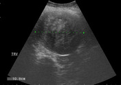
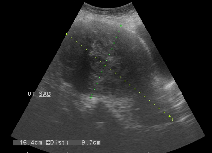
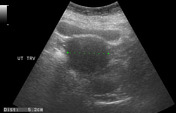
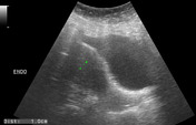
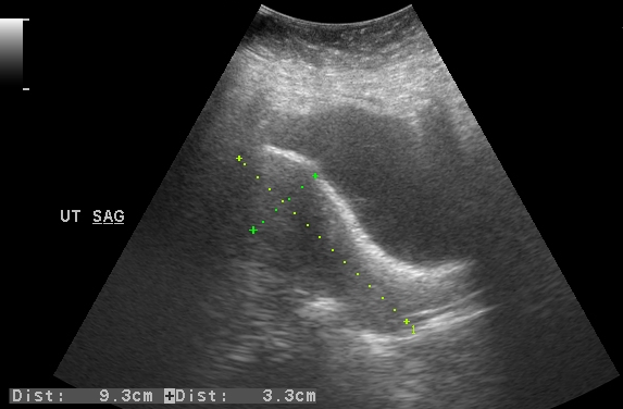
Bulky heterogeneous anteverted uterus with a heterogeneous structure measuring 9 x 9 cm noted in the fundal region. No cystic changes or calcifications are seen in it. No significant internal vascularity is seen in it.
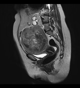

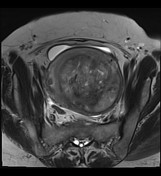
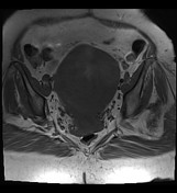
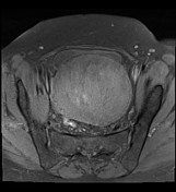
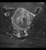

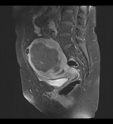
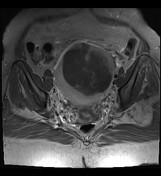

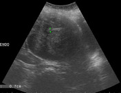
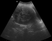
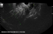
A heterogeneous, well-defined, slightly lobulated mass measuring about 11.0 x 9.5 cm, displacing the endometrium, is seen arising from the uterine myometrium. Multiple non-enhancing necrotic areas are seen in it. No extra-uterine extension or invasion of the surrounding structures is seen. No pelvic lymphadenopathy is noted. Ectopic left pelvic kidney with mild hydronephrosis.
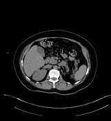
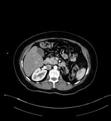
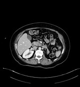
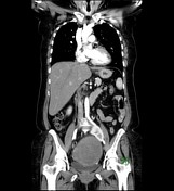
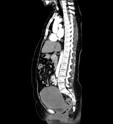
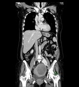
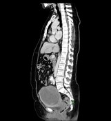
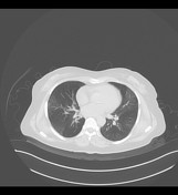
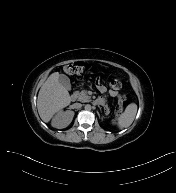
Large well defined uterine mass measuring 10 x 10 x 10 cm with central necrosis. No evidence of distant metastases is noted. Left pelvic kidney with mild hydronephrosis due to mass effect on the left ureter by the pelvic mass. Mosaic appearance of both lungs which is likely due to small airway disease. No suspicious pulmonary mass or nodule is seen.

Average size anteverted uterus with an endometrial stripe measuring 12 mm. No focal lesion/fibroid is seen in the uterus.
Case Discussion
Procedure: Total abdominal hysterectomy (TAH) and bilateral salpingo-oophorectomy (BSO).
Histopathology: Leiomyosarcoma. 11 cm in greatest dimension. Positive tumor cell necrosis. Myometrial invasion: focal less than 2 mm (myometrial thickness cannot be assessed). Mild to moderate focal atypia. Mitotic index=14 M/10 HPF. Endometrium, cervix, ovaries and fallopian tubes not involved. Negative margins. Pathologic staging: pT1b, pNx.


 Unable to process the form. Check for errors and try again.